

Custom Search
|

|
Soft
Tissue Pathology Pathology of Alveolar Soft Part Sarcoma Dr Sampurna Roy MD 2022
|

|
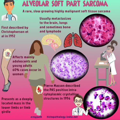
Alveolar soft part sarcoma (ASPS) is a slow growing but, highly malignant tumour. This rare, distinctive sarcoma, typically occurring in young patients was first described by Christopherson et al in 1952. Pierre Masson described the intracytoplasmic crystalline structures which stained positively with periodic acid‐Schiff stain in his 1956 text, Tumeurs humaines: histologie, diagnostics et techniques. Alveolar soft-part sarcoma: a review and update. ASPS is extremely rare, and is generally believed to account for less than 1% of all soft tissue malignancies Histogenesis of this tumour has not been established. Some have supported the idea that the tumour represents a distinct variant of rhabdomyosarcoma. However, subsequent studies have failed to demonstrate rhabdomyoblastic differentiation. Age and sex: Adolescents and young adults and rarely in children. 60% of ASPS cases are seen in women. Site: The tumour presents as a deeply located mass in the lower limbs or limb girdle. Rarely these are located in the trunk, retroperitoneum, head and neck. mediastinum and female genital tract. In children the tumour is usually located in the head and neck region (involve the tongue and orbit). Gross: Poorly circumscribed soft and friable tumour. Cut section reveals yellowish white to greyish red areas. Necrosis and haemorrhagic areas are usually present. Microscopic features- - Dense fibrous trabeculae divide the tumour into compartments of irregular sizes. - These are further divided into nests or islands of tumour cells. These islands are separated by thin walled vascular channels. - The tumour cells are large, round to oval shaped, with eosinophilic granular cytoplasm , eccentric rounded nuclei and prominent nucleoli. - The cellular aggregates show central degeneration and loss of cohesion, resulting in pseudoalveolar pattern. - Presence of PAS positive and diastase resistant intracytoplasmic granules and crystalline rods. - Pleomorphism may be present focally and mitotic figures are scarce. - Dilated veins are noted at the margin. Vascular invasion is common. - Rarely paraganglioma-like or pseudoglandular patterns and psammomatous calcification may be present. - In children the tumour is characterized by uniform sheets of large granular cells without nest-like arrangement. This variant has a better prognosis. Immunohistochemistry: An alveolar soft‐part sarcoma (ASPS) showing uniform, strong nuclear positivity with an antibody directed against the C‐terminus of transcription factor 3 (TFE3), confirming the presence of an ASPL–TFE3 fusion "Note: The diagnosis of alveolar soft part sarcoma is commonly based on characteristic histology and distinctive periodic acid-Schiff-positive crystals; however, the characteristic crystals may not always be observed, rendering the diagnosis difficult. Three important characteristics of alveolar soft part sarcoma, the presence of ASPSCR1-TFE3 fusion transcript, nuclear immunoreactivity for TFE3, and immunoreactivity for monocarboxylate transporter 1 and CD147, have recently been reported." Some cases stain positively with desmin, muscle specific actin, sarcomeric actin, S100 protein and neuron specific enolase. There is inconsistent demonstration of MyoD1. PAS positive diastase resistant cytoplasmic granules are strongly reactive to monocarboxylate transporter1 (MCT1) and CD147. Cytogenetics: Rearrangement of 17q 25 Gene expression profiling of alveolar soft-part sarcoma (ASPS) BMC Cancer. 2009;9:22. Electron micrograph, showing a typical intracytoplasmic crystal in an alveolar soft‐part sarcoma. Indicator of poor prognosis: (i) Older age (ii) Tumour size greater than 10 cms. Metastasis: The tumour usually metastasizes to the lungs and brain and sometimes to bones, lymph nodes. Brain metastasis is a common feature of Stage IV alveolar soft part sarcoma and routine intracranial imaging has been recommended as part of the staging evaluation in all patients with ASPS. The tumour may even metastasize 30 years after excision of the primary tumour. Differential diagnosis: - Metastatic renal cell carcinoma - Metastatic melanoma - Rhabdomyosarcoma - Paraganglioma - Metastatic adrenocortical carcinoma
- Malignant granular cell tumour " The differential diagnosis of ASPS involves a wide range of neoplasms that contain a similar alveolar nest-like pattern. This includes renal, adrenal and hepatocellular carcinomas, which have a similar eosinophilic cytoplasm, but are distinguished by cytokeratin markers. Malignant melanomas can be differentiated by PAS-D negativity and positivity for melanocytic markers. Unlike ASPS, paragangliomas show strong expression of neuroendocrine markers and S100 typically is positive in the sustentacular cells. Granular cell tumors are strongly positive for S100, and lack the characteristic vascularity of ASPS. Due to the alveolar architecture and occasional staining for muscle markers, a diagnosis of alveolar rhabdomyosarcoma may be considered, however, the cytology of ASPS is larger and with more abundant cytoplasm than seen in alveolar rhabdomyosarcomas. " Clavicular and meningeal alveolar soft part sarcoma: An unusual case and literature review.
|
|
|

![]()
Copyright © 2002-2022 histopathology-india.net