

Custom Search
|


|
Dermpath-India Pathology of Aneurysmal Variant of Dermatofibroma (Aneurysmal Fibrous Histiocytoma) Dr Sampurna Roy MD 2022
|
|
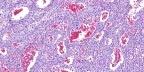
Aneurysmal benign fibrous
histiocytoma is an uncommon pathologic variant of dermatofibroma. In
addition to the features of a typical dermatofibroma, it has large
cleft-like or cavernous blood-filled spaces with numerous hemosiderin
pigments. Aneurysmal fibrous histiocytoma differ from the classical cutaneous fibrous histiocytoma in both their clinical presentation and pathologic features. Clinically, they may be larger than the usual cutaneous fibrous histiocytoma, are blue, black, or dark red, and have a cystic consistency. They are most commonly located on the extremities and may be associated with symptoms of pain and rapid growth. The clinical diagnosis of fibrous histiocytoma is rarely considered in the differential diagnosis, which may include malignant melanoma, hemangioma, neurofibroma, and nonspecific cyst. Histologically, the lesions are characterized by the presence of large, blood-filled tissue spaces. Most cases show degree of epidermal hyperplasia, as seen in common fibrous histiocytoma. These spaces lack an endothelial lining, being surrounded and lined by histiocytes, many of which contain hemosiderin pigment, fibroblasts, and foam cells. The solid portions of the tumor have the usual features of a cutaneous fibrous histiocytoma. Despite the presence of prominent secondary changes due to haemorrhage, many cases show cellular polymorphism, hyalinized collagen bundles surrounded by tumour cells in the periphery of the lesion. Immunohistochemistry: The tumour is usually positive for vimentin. There is rare focal smooth muscle actin positivity. CD68 is positive in some reactive macrophages only. Stains for CD31, CD34, desmin and factor XIIIa are usually negative. Differential diagnosis: 1) The presence of extravasated erythrocytes in combination with a spindle-cell stroma may lead to an erroneous diagnosis of Kaposi's Sarcoma. Kaposi's Sarcoma is positive for CD34 and lacks fibrohistiocytic cells. 2) Another important differential diagnosis is spindle cell angiosarcoma. Spindle cells in aneurysmal fibrous histiocytoma is negative for CD31 and CD34. Some histiocytes may be CD31 positive. 3) Malignant melanoma is excluded due to absence of an overlying in-situ component. Immunohistochemistry reveals that S100 protein is negative. 4) Angiomatoid fibrous histiocytoma, unlike aneurysmal dermatofibroma, tends to have cells that are more epithelioid compared to the spindle cells more commonly found in aneurysmal dermatofibroma. In addition, angiomatoid fibrous histiocytoma occurs exclusively in subcutaneous locations as opposed to aneurysmal dermatofibroma, which is situated predominantly in the dermis (with possible subcutaneous extension).
|
|
|
 |
