

Custom Search
|


|
Dermpath-India Pathology of Kaposi's Sarcoma Dr Sampurna Roy MD 2022
|
 |
|
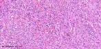
Kaposi's sarcoma (KS) is a low-grade, spindle-cell neoplasm
first described by Moritz Kaposi, in 1872. Although the exact pathogenesis of KS is not known, infection with HHV-8 / KS-associated herpes virus, combined with other genetic and environmental factors, has been strongly implicated as the cause of this disease. HHV-8 has been detected in KS cells of patients in all 4 epidemiologic groups, suggesting that the virus is a common pathogenetic factor for all KS types. (HHV-8 has also been implicated in the pathogenesis of multicentric Castleman's disease and primary effusion lymphomas.) HHV-8 infects CD19(+) B cells as well as T cells, monocytes, endothelial- derived spindle cells and CD34 (+) cells in the peripheral blood of patients with Kaposi's sarcoma. There are three subtypes of HHV-8 (A, B and C). Type C infection is usually not associated with extracutaneous disease. The tumour begins as a reactive proliferation but behaves as a multifocal neoplasm in advanced stage. The spindle cells in Kaposi's Sarcoma are thought to be the proliferating component while the endothelial cell population is thought to undergo a reactive hyperplasia. Some authors have suggested that the spindle cell elements show endothelial differentiation. Chronic stimulation of endothelial cells (possibly by viral infection) can produce differentiation to spindle shaped cells. Recent studies have indicated that the spindle cells are derived from the lymphatic endothelium. The Histogenesis of Kaposi's sarcoma cell: immunomorphological comparison with endothelial cells of normal human skin, lymphangioma and hemangioma. Arch Dermatol Res 1994;286:212 ; Expression of D2-40 in lymphatic endothelium of normal tissues and in vascular tumours.
|
| Kaposi's sarcoma can be
subdivided into 4 epidemiologic groups: 1. Classic variant: Usually affects elderly men of Eastern European and Mediterranean origin ; Usually present in fifth and seventh decade ; More common in men than in women, with a ratio 15 to 1 ; Presents as multiple firm, purple-blue or reddish-brown plaques and nodules ; Typically appear initially on the hands and feet ; Progress up the arms and legs over a period of years or decades; Involve the viscera or mucosa about 10 percent of patients ; Untreated lesions evolve from flat discolorations or patches to plaques and then to raised nodules that become confluent ; Histologic features - spindle-shaped tumour cells surrounding hyperemic vascular slits, extravasated erythrocytes, hemosiderin, and fibrosis ; Increased risk of lymphoma ; Homosexual men may be at increased risk for classic type Kaposi's sarcoma.
|
|
2. Epidemic (HIV- associated):
In 1981, Friedman-Kien et
al. described Kaposi's sarcoma involving lymph nodes, viscera, and
mucosa as well as skin in young homosexual men ;
This aggressive and frequently fatal epidemic variant of KS affected homosexual men with AIDS, 20 times as frequently as it did to male patients with hemophilia and AIDS who had similar degrees of immunosuppression ; This variant is also observed in intravenous drug users ; Epidemic variant of Kaposi's sarcoma has an extensive distribution and rapid progression.
|
| 3. Immunosuppression
associated: Present in
organ-transplant
recipients and patients who are receiving corticosteroid or immunosuppressive
therapy for a variety of medical conditions ;
This type of Kaposi's sarcoma tends to be aggressive,involving lymph nodes, mucosa, and visceral organs in about half of patients, sometimes in the absence of skin lesions ; The presence of concurrent lymphoma, tuberculosis, or transfusion-related HIV infection makes it difficult to diagnose KS accurately.
|
| 4. African (endemic) :
In the 1950s, KS was recognized as being common
in portions of Africa. It was reported in
Uganda and Zambia.
In eastern and southern Africa, KS makes up 25 to 50 percent of
soft-tissue sarcomas in children. Subtypes: Lymphadenopathic type: Usually noted in children. The patients have a poor prognosis. Nodular disease: Resembles classic type. Aggressive atypical variant : Characterized by generalized, infiltrative skin lesion in adults. This variant responded poorly to conventional treatment.
|
|
Histopathological features:
Patch ; plaque ; nodular ; lymphadenopathic ; infiltrative ; florid ; telangiectatic ; ecchymotic ; keloidal ; angiomatous ; inflammatory ; anaplastic ; lymphangiomatous ; and generalized lymphedema . Patch Stage: Flat lesion characterized by proliferation of numerous jagged vascular spaces in the dermis ; The vascular spaces are parallel to the epidermis ; The slit like vessels are present around preexisting blood vessel , skin adnexa and between collagen fibres. ( 'Promontory sign' ) ; Vessels are lined by plump, mildly atypical endothelial cells ; Perivascular lymphocytes and plasma cells ; Extravasated red blood cells and hemosiderin may be present ;
The features resemble granulation tissue. Spindle cells are more prominent than those in the 'patch stage' ; Dermal proliferation of the spindle cells together with poorly defined slit-like blood vessels ; Involves the reticular dermis and even the subcutis ; Hemosiderin deposition is prominent ; Eosinophilic globules are present . Nodular Stage: Well defined lesion characterized by prominent interlacing bundles of spindle cells around slit like blood vessels and extravasation of red blood cells ; These features are more prominent than those in the 'plaque stage' ; Dilated thin walled vessels are present at the periphery ; Mitotic figures are present ; Eosinophilic hyaline globules are present (intra and extra cellular) ; These are PAS- positive and stain bright red with Mallory's trichrome. Lymphangioma-like Kaposi's Sarcoma (LLKS): Clinically, the lesions have a bulla like appearance. Histologically, the tumour is characterized by permeation of dermal collagen by irregular anastomosing vascular channels lined by a flattened endothelium. The biopsy specimens also reveal areas with characteristic light microscopic features of KS. All tumour cells, show a strong and diffuse reactivity for anti-HHV-8 LNA-1 and anti-CD34. Anaplastic variant of Kaposi's Sarcoma: Cases have been reported in Africa. Characterized by : Greater cellularity ; Nuclear pleomorphism ; Frequent mitotic activity.Kaposi's sarcoma: histopathological study of 159 cases from Malawi; Kaposi's sarcoma in Uganda: a clinico-pathological study. Immunohistochemistry: Kaposi's sarcoma is positive for both CD31 and CD34. Some cases are positive for factor VIII-related antigen. Human herpes virus type - 8 can be detected by PCR in paraffin-embedded tissue. Recently, a monoclonal antibody to human herpes virus 8 latent nuclear antigen-1 has become commercially available for immunohistochemical analysis. Kaposi sarcoma shows strong, diffuse, nuclear staining for human herpes virus 8 latent nuclear antigen-1. Differential Diagnosis of Kaposi's Sarcoma: Early vascular lesions should be differentiated from telangiectasia, pigmented purpuric dermatosis , acroangiodermatitis (Acro-angiodermatitis. A simulant of Kaposi's sarcoma. ) and low grade angiosarcoma. Vessels in KS are more irregular. An inflammatory infiltrate which includes plasma cells is present in early lesions of KS. Regressed KS lesions following therapy may be misdiagnosed clinically and histologically as pigmented purpuric dermatitis if the pathologist is not aware of the previous history. The histologic differential diagnoses of KS include other vascular tumours, such as spindle cell hemangioendothelioma , kaposiform hemangioendothelioma, and angiosarcoma. Spindle cell hemangioendothelioma: In addition to a solid spindle cell component resembling KS, there are dilated and cavernous vascular spaces. Kaposiform hemangioendothelioma is a pediatric tumour composed of multiple lobules, each of which resemble either KS or capillary hemangioma, and is often associated with Kasabach-Merritt syndrome. HHV-8 has not been associated with kaposiform hemangioendothelioma. Angiosarcoma is characterized by a complex anastomosing pattern and marked nuclear atypia. The presence of hyaline bodies and deposits of haemosiderin indicate Kaposi's sarcoma. The spindle cell predominant type KS may be confused with leiomyoma, leiomyosarcoma, or fibrosarcoma.
The presence of hyaline bodies and the
formation of vascular channels between spindle cells point to a diagnosis
of Kaposi's sarcoma. Other histologic simulators of Kaposi's sarcoma are: -Intranodal Palisaded Myofibroblastoma; -reactive angioendotheliomatosis ; -aneurysmal fibrous histiocytoma ; -nonspecific vascular proliferation; -angiomatoid fibrous histiocytoma ; -dermatofibrosarcoma protuberans; -vascular transformation of lymph node ; -pilar leiomyoma ( Piloleiomyoma ) ; -stasis dermatitis ; -spindled melanoma. Dermatopathology Quiz Case 142
|
|
|
|
|

![]()
Copyright © 2002-2022 histopathology-india.nett