Custom Search


Dermpath-India

Pathology of Clear Cell Acanthoma  

"Skin lesion with enlarged pale stained keratinocytes" 

Dr Sampurna Roy MD        2022


Dermatopathology Quiz Case: 149

Answer -    Clear Cell Acanthoma

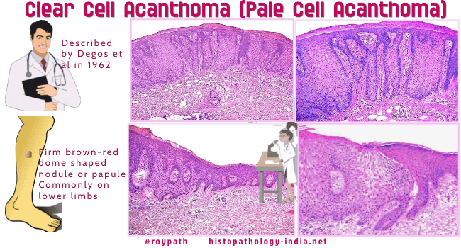
Clear cell acanthoma (pale cell acanthoma) is a discrete idiopathic psoriasiform lesion of large pale staining, non-keratinizing, glycogenated, monomorphous keratinocytes associated with neutrophils. The lesion is separated from the adjacent epidermis.

(Dermatopathology Poster )

It was first described by Degos et al in 1962.

Clinical presentation:  Presents as a firm brown-red dome shaped nodule or papule.

The lesion is usually between 5 to 10 mm or more in diameter. 

Clear cell acanthoma is usually solitary but can be multiple.

Clinical bleeding is rare.

The surface of the lesion may be smooth to bosellated and crusted.

Age:  Middle aged and elderly individuals. Some cases have been reported in younger patients.

Site: Commonly located on the lower limbs. These may also be found on the trunk and face.


Microscopic features:  

Well-demarcated and symmetric lesion elevated in comparison to surrounding skin.

The base of the lesion situated below the rete-ridges of the normal epidermis.

A sharp line can be drawn between the lesional cells and normal epidermis.

There is psoriasiform hyperplasia of the rete ridges with broad base.

The keratinocytes have pale-staining cytoplasm.

A PAS stain with and without diastase confirm the presence of abundant glycogen in the cells.

There is usually no cellular atypia.

Other epidermal changes include mild spongiosis and exocytosis of neutrophils forming small intraepidermal microabscesses.

The epidermal surface shows parakeratotic scales.

Melanocytes within the lesions are variable in number. 

A pigmented variant has been reported.

Papillary dermis is edematous and dermal capillaries are prominent.

An inflammatory infiltrate composed of lymphocytes , plasma cells and rarely eosinophils are present.


Immunohistochemistry:  The cells contain cytokeratin and involucrin but not carcinoembryonic antigen.

Differential diagnosis: Eccrine poroma ; Trichilemmoma ; Psoriasis; Clonal type-Seborrheic keratosis ; Verruca vulgaris  

Dermatopathology Case No 25


Further reading:

Clear cell acanthoma with changes of eccrine syringofibroadenoma: reactive change or clue to etiology?

Multiple clear-cell acanthomas.

Clinicopathologic and immunohistochemical studies of conjunctival large cell acanthoma, epidermoid dysplasia, and squamous papilloma.

Clear cell acanthoma of the areola and nipple: clinical, histopathological, and immunohistochemical features of two Brazilian cases.

Clear cell acanthoma.

Clear cell acanthoma induced by a dermatofibroma.

Hemosiderotic clear-cell acanthoma: a pigmented mimicker.

Clear cell acanthoma: a rare clinical diagnosis prior to biopsy.

A case of polypoid clear cell acanthoma on the nipple.

Clear (pale) cell acanthosis as an incidental finding.

A case of polypoid clear cell acanthoma on the scrotum.

Polypoid clear cell acanthoma of the scalp.

Clear cell acanthoma presenting as polypoid papule combined with melanocytic nevus.


Visit:- Infectious Disease Online


Dr Sampurna Roy  MD

Consultant  Histopathologist (Kolkata - India)


Copyright © 2022  histopathology-india.net