

Custom Search
|

|
Infectious Disease Online Pathology of Chagas' Disease (American Trypanosomiasis)
|
| Syn: Kissing Bug
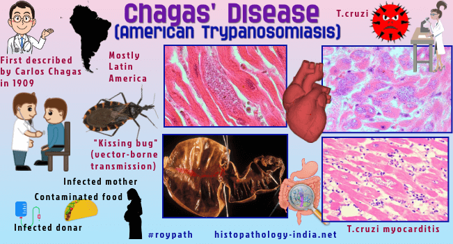
Disease Chagas disease, also known as American trypanosomiasis, is a potentially life-threatening illness caused by the protozoan parasite Trypanosoma cruzi (T. cruzi). Trypanosoma cruzi, causes acute, subacute and chronic parasitemia, with dissemination to many organs, specially the heart, brain, esophagus and colon. Chagas disease is named after Carlos Ribeiro Justiniano Chagas, a Brazilian physician and researcher who discovered the disease in 1909. Most T. cruzi infections are mostly transmitted to humans by contact with faeces or urine of triatomine bugs (vector-borne transmission), known as 'kissing bugs'. Other modes of transmission include transfusion of contaminated blood products, organ transplantation, congenital transmission, laboratory accidents, and the ingestion of contaminated food and drink. Trypanosoma cruzi occurs in blood as a trypomastigote and in reticuloendothelial and other tissue cells typically as a leishmanial form, the amastigote. The amastigote form lacks flagella but retains the kinetoplast, which permits its differentiation from other intracellular organisms such as Toxoplasma and Histoplasma. The disease occurs in North and South America, mostly in the area from Mexico to Argentina. It has never been reported outside the Western Hemisphere, though the vectors and animal reservoirs are common to many parts of the world. The vectors are large biting insects of the genus Triatoma.These insects are found in areas where there are unhygienic conditions associated with poverty. They bite only at night. During the day they hide in cracks in the walls of primitive country dwellings. The organism is sucked up by the Reduviids, "the kissing bugs", as a free flagellate or as an intracellular leishmanial form within a macrophage. In the midgut of the insect the organism becomes flagellated, and binary multiplication occurs. It migrates to the hindgut, where it is transformed into the metacyclic trypanosome form infective for the vertebrate host. Since the triatoma defaecates at the time of biting, the infection is usually acquired by rubbing the faeces containing the metacyclic trypanosome stage of the parasite into the tiny skin puncture or into other abrasions of the skin in the area. Infections through the intact mucous membranes occur most frequently in the lips or in the conjunctivae (when the eyes are rubbed with fingers soiled with the insect faeces) . Trypanosoma cruzi enters macrophages by parasite-specified phagocytosis. Phagocytosis is partially dependent on macrophage cell surface receptors. The protozoon does not remain within phagocytic vacuole but escapes into the cytoplasm where it multiplies. By leaving the parasitophorous vacuole, the organism escapes the action of microbicidal lysosomal enzymes. The circulating forms are susceptible to the trypanosomal activity of the lysosomal enzymes and major basic protein of eosinophils. Specific diagnosis of America trypanosomiasis may be accomplished by the finding of the typical trypanosome stage of Trypanosoma cruzi in the blood during febrile episodes. Aspiration of spleen, liver, lymph nodes, or bone marrow will frequently reveal the leishmanial form of the organism in fixed macrophages. In reticuloendothelial cells Trypanosoma cruzi cannot be easily distinguished from species of leishmania, but only Trypanosoma cruzi invades myocardial and neuroglial cells as a leishmanial type of organism. Serologic methods include complement fixation, indirect immuno-fluorescence, hemagglutination, enzyme-linked immunosorbent assay and thin layer immunoassay. Acute Chagas' Disease: The acute form is the more common of the two types of Chagas' disease. It occurs predominantly in small children and is characterized by moderate hepatosplenomegaly, facial edema, tachycardia, and the presence of T. cruzi in peripheral blood. In 50% of cases the primary site is the outer canthus of one eye, with unilateral palpebral edema and satellite preauricular lymph node enlargement (Romana's sign). In 25% of cases the portal of entry is represented by a nodular or ulcerative skin lesion (chagoma), accompanied by enlargement of regional lymph nodes. Histologically, the primary lesion is characterized by numerous histiocytes, chronic inflammatory cells, and areas of fat necrosis throughout which numerous leishmanial forms are identified. Multiplication of the organisms that have been engulfed by nearby macrophages is soon followed by the invasion of other structures such as smooth and striated muscle (including cardiac muscle), glial and nervous cells and fat cells. Parasites are found in almost all organs, and their presence is accompanied by mononuclear cell infiltration, congestion, and edema, and occasionally by granulomatous areas. Reticuloendothelial activity and increase in the number of fixed macrophage cells cause splenomegaly, hepatomegaly, adenopathy and hyperplasia of bone marrow. The process is then a reticulopathy similar to kala-azar and histoplasmosis. Death during acute phase is caused by acute myocarditis with congestive cardiac failure, meningoencephalitis, or complications such as bronchopneumonia. A recent study showed that T. cruzi readily enters the central nervous system during the acute infection. Cerebrospinal fluid findings include pleocytosis and high protein levels. The heart is enlarged and flabby with pronounced dilatation of the left ventricle. The myocardium is pink and shows yellowish gray streaks. Microscopically, the myocardium is diffusely involved but reveals only spotty cell destruction. The myocardial fibers are separated by intense edema, proliferation of histiocytes, and infiltration with chronic inflammatory cells and few polymorphonuclar cells. Within the muscle fibers there are leishmanial forms either arranged in rows or in cyst-like dilatations. Cardiac fibers show degenerative changes, the most important of which is hyaline necrosis of isolated fibers. Inflammatory changes in the myocardium also may involve the bundle of His and its ramifications. Chronic Chagas' Disease: The dominant factor in the chronic stages of Chagas' disease is almost always some degree of myocardial fibrosis. Many patients report a previous acute attack. However, in a number of cases there is no history of a previous attack. The chronic cardiac form of Chagas' disease is a leading cause of cardiac failure and sudden death in endemic areas. The heart is enlarged with generalized hypertrophy and dilatation. Mural endocardial thromboses are usually present, most frequently in the right auricle and at the apex of the left ventricle. Apical thrombosis, with focal endomyocardial fibrosis at the base, represents the most important single gross finding suggestive of Chagas' myocarditis. In the absence of thrombosis the myocardium at the apex frequently appears distended and thinned, sometimes even to the point of aneurysmal formation. Embolic phenomena, both pulmonary and systemic are present in about 75% of autopsied cases. The main source of emboli are intracardiac thrombi which are also found at autopsy in approximately 75% of cases. Microscopically the myocardium shows chronic, non-specific, mononuclear inflammation. Hypertrophy of cardiac fibers, small focal areas of necrosis and granular degeneration of cardiac fibers, focal and diffuse fibrosis, vascular dilatation and congestion, and interstitial and interfibrillary edema are other common microscopic features. Parasites (leishmanial forms in pseudocysts) are identified with difficulty in a very limited percentage of cases. Thus the pathogenesis of myocardial fiber damage in chronic Chagas' disease is still unclear. Two primary mechanisms, parasite-driven immune response and autoreactivity triggered by the infection, likely initiate and drive both acute and chronic myocarditis, with 2 secondary mechanisms, neurogenic disturbances and coronary microvascular derangements, being responsible for associated cardiac alterations.Pathogenic differences in T cruzi strains and host susceptibility also likely play a role in the clinical pattern and disease severity. Gastrointestinal disease of Chagas' Disease: Chagas gastrointestinal disease is the result of enteric nervous system impairment, creating disordered esophageal or colonic motility. Esophageal involvement ranges from mild achalasia to severe megaesophagus, characterized by dysphagia, odynophagia, esophageal reflux, weight loss, aspiration, cough, regurgitation, and increased risk of esophageal carcinoma.Colonic involvement typically manifests as constipation with megacolon, which can be complicated by fecaloma, volvulus, and bowel ischemia. Fibrosis and degeneration of autonomic ganglia in the heart have been demonstrated in persons with megaesophagus, as well as considerable diminution in the number of ganglionic cells in the Auerbach plexus of the esophagus and intestine. The prognosis for patients with digestive forms of the disease is generally good except in those with advanced forms and complications that occasionally cause death Congenital Chagas' Disease: The transplacental transmission of T. cruzi infection has been well documented in humans. Congenital Chagas' disease can lead to premature birth birth and fetal death. A chronic placentitis, similar in some respects to the syphilitic placenta, with bulky and ischemic villi, has been described. Leishmanial forms are found within the cytoplasm of macrophages in variable numbers. Interstitial pneumonitis with a prominent mononuclear histiocytic infiltrate can develop in infants.
|
|
|
