Custom Search


Dermpath-India

Pathology of Dilated Pore of Winer

Dr Sampurna Roy MD               2022
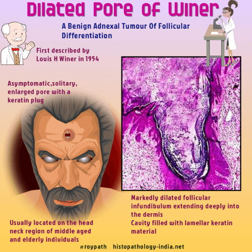
 

Dermatopathology Quiz Case: 263

Answer -Dilated Pore of Winer 


Dilated pore of Winer is relatively common but poorly recognized adnexal lesion. The Winer dilated pore.Med Cutan Ibero Lat Am. 1989;17 (1):45-7.

The lesion is commonly noted in elderly patients.

Clinically the lesion looks like a giant comedo.

These are usually located in the head, neck and upper trunk.


Microscopic features:

- Lesion may extend to mid or lower dermis ;

- Funnel shaped structure lined by infundibular or isthmic-type epithelium,

- It is filled with laminated keratin, connected to the surface ; 

- Thickening of epithelium including finger-like projection into the dermis ;

- Epidermoid keratinization.


Differential diagnosis: Pilar sheath acanthoma


Further reading:

Dermoscopy of a dilated pore of Winer.

Hair cortex comedo: a series of 34 cases.

Winer's dilated pore of the eyelid.

Dilated pore: a case report and an immunohistochemical study of cytokeratin expression.

Dilated pore of Winer.

Trichoid basal cell carcinoma found in a dilated pore on the nose.

The Winer dilated pore.


Visit:- Infectious Disease Online

Dr Sampurna Roy  MD

Consultant  Histopathologist (Kolkata - India)


Copyright © 2002-2022  histopathology-india.net