Custom Search
|
|
Custom Search
|
|
Dermpath-India Pathology of Pilar Sheath Acanthoma Dr Sampurna Roy MD 2022
|
|
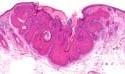
Pilar sheath acanthoma is a rare, benign follicular hamartoma described first in 1978 by Mehregan and Brownstein. Clinical presentation: Clinically resembles a comedo and is characterized by a central pore - like opening plugged with keratin. Lesion is 5-10mm in diameter. Site: Solitary lesion occuring almost always on the upper lip. Cases have been reported on other areas of the head and face.
Differential diagnosis: 1. Dilated pore (small finger-like projections extending into the dermis) ; 2. Trichofolliculoma (secondary hair follicles radiate from the wall of the primary follicle, an outer root sheath, inner root sheath and trichohyaline granules are noted in the secondary follicles) ; 3. Poroma
|
|
|
 |
Visit:-
Infectious Disease Online

Consultant Histopathologist (Kolkata - India)
|
![]()
Copyright © 2002-2022 histopathology-india.net