

Custom Search
|


|
Dermpath-India Pathology of Giant Cell Collagenoma
|
|
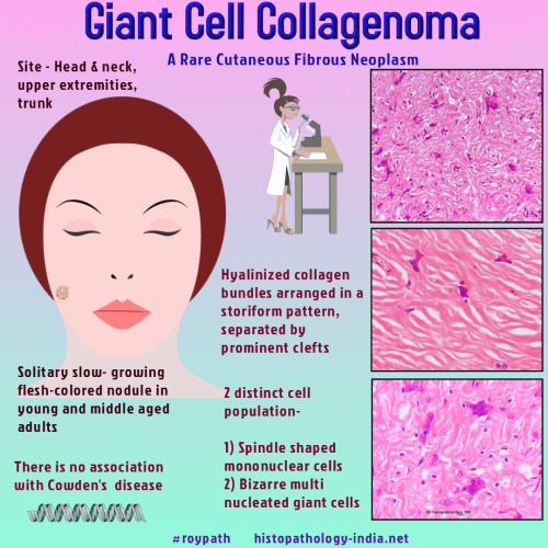
Giant cell collagenoma was described for the first time by Rudolph et al in 1998. It is a cutaneous fibrous neoplasm that usually affects young to middle-aged adults. Despite its similar histological appearance with circumscribed storiform collagenoma, no association of Giant cell collagenoma with Cowden's syndrome has been described so far. Gross: Presents as solitary slow-growing flesh-coloured nodules in young and middle-aged adults. Microscopic feature: These lesions are characterized by sharply demarcated matrix consisting of coarse hyalinized collagen bundles arranged in a prominent storiform pattern and separated by mucin-containing clefts. Admixed with the collagen matrix, there are two distinct cell populations. (i) spindle-shaped mononuclear cells, and (ii) bizarre multinucleated giant cells. Atypical nuclei or mitotic figures are not present. Immunohistochemistry: Mononuclear cells expressed vimentin and actin HHF35. Multinucleated cells only expressed vimentin.
The cells are negative
for cytokeratin, desmin, S-100 protein, CD34, factor XIIIa,
and the macrophage markers KP1, Mac 387.
|
|
|
