Custom Search
|
|
Custom Search
|
|
Dermpath-India Pathology of Storiform Collagenoma (Sclerotic Fibroma)
|
|
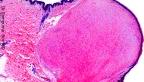
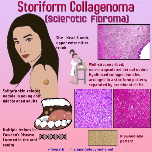
Storiform collagenoma or sclerotic fibroma is a rare benign skin neoplasm that usually affects young and middle-aged adults. This lesion was first described in association with Cowden's disease by Weary et al.in 1972. In 1989, Rapini and Golitz described 11 cases of solitary sclerotic fibroma in the absence of Cowden's disease, suggesting the term 'solitary sclerotic fibroma' of the skin. The terms hypocellular fibroma and circumscribed storiform collagenoma have also been used for this entity. Site: This tumor is often found in the head and neck region, upper extremities and chest. Multiple lesions are usually located in the oral cavity. Gross: They are small white or flesh-colored waxy papules ranging in size from 0.5 to 1.2 cm. Multiple lesions of this type have been reported in the multiple hamartoma syndrome (Cowden's disease). Microscopic features: These are well-circumscribed, unencapsulated dermal nodules characterized by epidermal atrophy, a whorled appearance of sclerotic collagen bundles separated by clefts containing mucin, and sharp demarcation of the lesions from the surrounding normal skin. The hyaline collagen bands are virtually acellular. Immunohistochemistry: The tumour cells express CD34, vimentin and factor XIIIa, but are negative for markers of neural or melanocytic differentiation. In some cases alpha-smooth-muscle actin positive myofibroblasts are present. Differential Diagnosis : Sclerotic fibroma shares a common immunoprofile with Pleomorphic Fibroma (discussed later), but is distinct from dermatofibroma (Sclerotic variant of dermatofibroma) and other common spindle cell lesions of skin. Pacinian collagenoma: Composed of paucicellular collagen fibres arranged in concentric lamellations giving rise to an onion-skin appearance. The cells are CD34 positive. Differential diagnosis: Perineurioma Pleomorphic sclerotic fibroma: In the superficial portion the tumour showed features of a pleomorphic fibroma, the deeper portion showed features of a sclerotic fibroma, and a transitional area is present in between. According to some authors, pleomorphic fibroma, sclerotic fibroma, and pleomorphic sclerotic fibroma form a spectrum. |
|
|
Visit :- Giant Cell Collagenoma
Visit:- Infectious Disease Online
![]()
Copyright © 2022 histopathology-india.net