Custom Search
|
|
Custom Search
|
|
Dermpath-India Pathology of Gouty Tophus Dr Sampurna Roy MD 2022
|
 |
|
Hyperuricemia causes urates to be deposited in the skin and sometimes
in the subcutaneous fat.
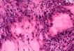
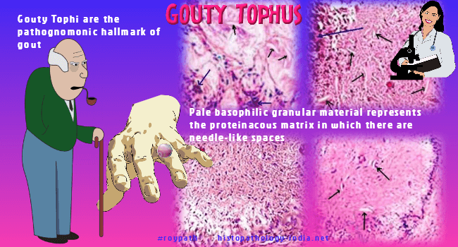
Gouty tophi are the pathognomonic hallmark of gout. They are formed by large aggregations of urate crystals surrounded by an intense inflammatory reaction of macrophages,lymphocytes and large foreign body giant cells which may have completely or partially engulfed mass of crystals. Tophi may appear in the articular cartilage of joints and also in the periarticular ligaments, tendons, soft tissues including the olecranon and patellar bursae, Achilles tendons, and ear lobes. Less frequently they may appear in the kidneys, nasal cartilages, skin of the fingertips, palms and sole. Superficial tophi can lead to large ulcerations of the overlying skin. Histopathological features: Pale basophilic granular material represents the proteinaceous matrix in which there are needle-like spaces in radial arrangement. These spaces represent outlines of urate crystals that have been dissolved by aqueous fixative. The pale basophilic area is surrounded by abundant histiocytes and foreign body type of giant cells. Note: Fixation in alcohol is important for the preservation of sodium urate monohydrate deposits that appear as needle-shaped, doubly refractile crystals. With De Galantha's stain, urate crystals appear brown-black. Differential diagnosis: - Rheumatoid nodule: Palisading of histiocytes may be a source of confusion with rheumatoid nodule. - Chondrocalcinosis (pseudogout). - Calcinosis Cutis: Calcified nodules of scrotum or vulva.
|
|
|
|
|
Visit:- Infectious Disease Online
![]()
Copyright © 2002-2022 histopathology-india.net