

Custom Search
|


|
Dermpath-India Pathology of Microvenular Hemangioma Dr Sampurna Roy MD 2022
|
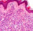
Microvenular hemangioma is a rare, slowly growing, benign vascular tumour. Clinical presentation: It usually presents as a solitary, asymptomatic, purple to red papule or plaque. Age: Young to middle-aged adults. Site: Extremities, particularly the forearms. Microscopic features: Histologically, these tumours are characterized by a proliferation of small-sized, irregularly branched venules with inconspicuous lumina. The tumour has an infiltrative growth throughout the dermis. The backround stroma is desmoplastic. The flattened endothelial cells have oval to spindle-shaped nuclei and scant cytoplasm. The endothelial cells are surrounded by pericytes. The tumour cells lack cellular atypia, pleomorphism, and mitotic figures. Immunohistochemistry: Endothelial cells: FactorVIII- related antigen and CD34 positive. Pericytes: Smooth muscle actin positive. Differential diagnosis: Early onset Kaposi's sarcoma- Eosinophilic globules are not present in microvenular hemangioma. In the context of the histological similarity to a low-grade malignant tumour - early onset Kaposi's sarcoma, the awareness of microvenular hemangioma, a benign vascular tumour, is important.
|
|
|
 |
