Custom Search
|


|
Dermpath-India Pathology of Necrobiotic Xanthogranuloma Dr Sampurna Roy MD 2022
|
|
Custom Search
|
|
Dermpath-India Pathology of Necrobiotic Xanthogranuloma Dr Sampurna Roy MD 2022
|
|
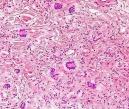
Necrobiotic xanthogranuloma is a rare disorder which is frequently associated with paraproteinemia and lymphoproliferative diseases. Age: Middle-aged or elderly patients. Clinical presentation: Lesion presents as reddish, partly xanthomatous nodules or plaques. Site:
These are usually located around the periorbital area. Other sites include
extremities and trunk.
Differential diagnosis: Necrobiosis Lipoidica: Much less collagen degeneration. Foam cells, large numbers of necrotic inflammatory cells and cholesterol clefts are absent. Atypical giant cells are not prominent. Panniculitis is localized to the septa. In necrobiotic xanthogranuloma the lobules are extensively involved.
|
|
|
 |
Visit:- Infectious Disease Online
![]()
Copyright © 2002-2022 histopathology-india.net