

Custom Search
|


|
Dermpath-India Pathology of Pleomorphic Hyalinizing Angiectatic Tumour Dr Sampurna Roy MD 2022
|
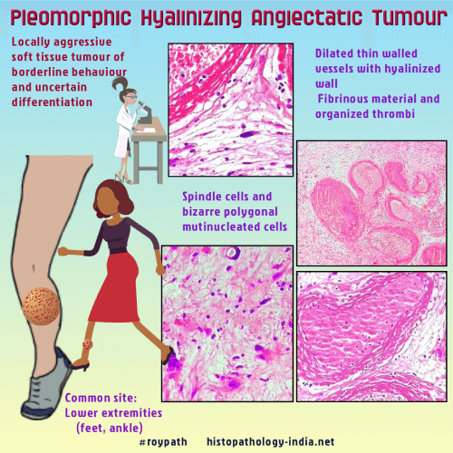
Syn: Pleomorphic Hyalinizing Angiectatic Tumour of Soft Parts Pleomorphic hyalinizing angiectatic tumour is a rare tumour of uncertain lineage. It was first described in 1996 in a series of 14 cases. It is a low-grade sarcoma which may recur locally. Metastasis has not been recorded. Site: The tumour is seen in the subcutaneous tissue of the distal extremities in adults. It is commonly noted on the ankle and feet. Gross: These are circumscribed or infiltrative lesion. Most tumours have a lobulated appearance with a variegated, tan-brown hemorrhagic cut surface. Cystic and myxoid changes may be present. Pleomorphic Hyalinizing Angiectatic Tumour [Pathology Infographic] Microscopic features: 1. Clusters of dilated thin-walled vessels with fibrinoid change of their wall with superimposed thrombosis & occasionally hemosiderin-laden macrophage. 2. Intervening stroma contain spindle shaped cells and bizarre polygonal multinucleate cells. These multinucleate cells show marked nuclear pleomorphism, hyperchromasia and frequently intranuclear inclusions. In rare cases psammomatous calcifications may be noted. 3. A variable inflammatory background mainly composed of lymphocytes, plasma cells, and numerous mast cells. Immunohistochemistry: Multinucleate cells are CD34 positive (50% cases) but negative for S-100 protein, actin and desmin. Differential diagnosis: 1. Ancient schwannoma (distinguished from schwannomas by the usual presence of infiltrative margins and the absence of S-100 protein) ; 2. Melanotic schwannoma (presence of hemosiderin, psammomatous calcifications, and intranuclear inclusions). 3. Primary cutaneous myxofibrosarcoma (Primary cutaneous myxofibrosarcoma mimicking pleomorphic hyalinizing angiectatic tumor (PHAT): a potential diagnostic pitfall.Am J Dermatopathol. 2005 Aug;27(4):322-6.) 4. Hemosiderotic fibrohistiocytic lipomatous lesion: Abstract 5. Some early cases may be mistaken for spindle cell lipoma, benign fibrous histiocytoma or nodular fasciitis. Local recurrence is common in almost 50% of cases. |
|
|
