

Custom Search
|


|
Dermpath-India Pathology of Proliferating Trichilemmal Tumour Dr Sampurna Roy MD 2022
|
 |
|
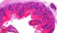
Trichilemmal (pilar) cysts are common skin lesions that usually occur on the scalp of elderly women. They differentiate towards the follicular outer root sheath epithelium and show trichilemmal keratinization. Proliferating trichilemmal tumour shows features of typical pilar cyst, but additionally shows extensive epithelial proliferation, variable cytologic atypia and mitotic activity. Proliferating trichilemmal tumour arises from the isthmus region of the outer root sheath. Clinical presentation: Usually occurs in elderly women and presents as a slowly enlarging, painful subcutaneous scalp nodule. Sites: Scalp is the most common site. Other sites include forehead, nose, back, chest, abdomen, buttocks, elbow, wrist, and vulva . Gross: Well defined and lobulated lesion (0.4 to 10 cm in diameter).
Differential diagnosis : Squamous cell carcinoma - (Proliferating trichilemmal tumour is characterized by abrupt keratinization, minimal pleomorphism, low mitotic activity, sharp circumscription, foci indistinguishable from a trichilemmal cyst, calcification and absence of a premalignant lesion such as actinic keratosis). The tumour often
recurs if not fully excised. Malignant Proliferating Trichilemmal Tumour:
This malignant tumour originates from a
preexisting trichilemmal cyst. Patient complains of sudden rapid growth . Site: Confined to the scalp, back or neck. Microscopic features: - Areas of transformation from benign proliferation of pilar sheath epithelium to an obvious malignant neoplasm ; - It is characterised by poorly defined borders, clear cut infiltrative properties with cytological evidence of malignancy ; - Extensive dermal lobular proliferation of pilar sheath epithelium is seen together with palisading arrangement of nuclei at the periphery of lobes ; - Multiple central areas of trichilemmal keratinization with occasional areas of calcification and homogeneous keratin cyst formation ; - Epithelium shows severe disarrangement of epithelial cells with large number of cells having irregular shaped hyperchromatic nuclei ; - There is brisk mitosis ; dyskeratotic cells ; shadow cells and multinucleated cells are also present ; - Tumour invades into the surrounding connective tissue.
|
