

Custom Search
|

|
Infectious Disease Online Pathology of Protothecosis
|
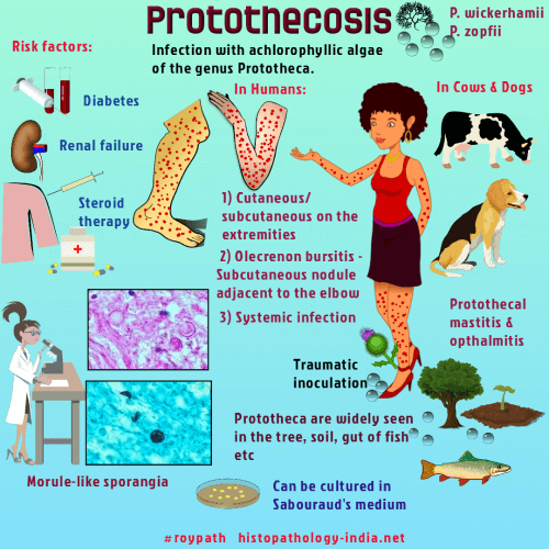
Protothecosis is an infection caused by achlorophyllous algae of the genus Prototheca. Although they do not contain chloroplasts, these saprophytic algae are believed to be related to green algae of the genus Chlorella. Causative organism: Four species of protothecae are recognized, of which two, Prototheca wickerhamii and Prototheca zopfii, are known to cause disease. Almost all authenticated cases of human protothecosis have been caused by Prototheca wickerhamii. Source of infection: The source of infection is often not apparent but can be related to penetrating injury in some cases. Clinical forms: Three clinically distinct forms of protothecosis are recognized: 1) Cutaneous protothecosis, which occurs preferentially in debilitated or compromised patients, present as spreading papulonodular or verrucous lesions, usually involving the distal extremities or head. Infection may extend into the subcutaneous tissue and rarely spreads to regional lymph nodes. 2) Olecranon bursitis, which occurs in otherwise healthy hosts, presents as a subcutaneous nodule adjacent to the elbow. Bursectomy is the treatment of choice. 3) Systemic infection.
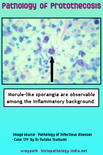
Chemotherapy alone is not effective in eradicating localized infection in most cases. The single reported case of disseminated human protothecosis occurred in a patient with transient depression of specific cell-mediated immunity to Prototheca. He recovered after therapy with transfer factor and amphotericin B. Life cycle and features of the two species: The protothecae are found in tissue sections in the form of endosporulating sporangia. Their asexual reproductive cycle in tissue is similar to that of the endosporulating fungi. Small, uninucleate, immature sporangia undergo nuclear division followed or accompanied by progressive cytoplasmic cleavage to produce mature sporangia that contain sporangiospores. Characteristically, the sporangiospores are polygonal or wedge-shaped, fill the parent cell, and may be radially arranged around a central sporangiospore, producing the distinctive “morula” form. The sporangia of the two pathogenic protothecae differ in size but are otherwise similar in morphology. Sporangia of the small form, Prototheca wickerhamii, measures 2 to 12 micrometer in diameter, where as those of Prototheca zopfii measures 10 to 25 micrometer in diameter. Morula forms are uncommon in infections caused by Prototheca zopfii. Endosporutating cells of Prototheca zopfii are oval, and their larger nuclei are more conspicuous than those of Prototheca wickerhamii. The cell walls of both the sporangia and the sporangiospores are stained with the special stain for fungi. With hematoxylin and eosin, these cells are hyaline, but their contents may be eosinophilic or basophilic. The two species are more reliably distinguished from one another in tissue sections by direct immunoflourescence and in culture by their patterns of carbohydrate assimilation.
Pathological features: Cutaneous lesions often show hyperkeratosis, parakeratosis, and acanthosis, and they may be ulcerated. Algal cells are abundant in the dermis and may also be found in the epidermis and keratin layer as a result of transepidermal elimination. An inflammatory reaction, when present, may be granulomatous or may consist of a mixture of acute and chronic inflammatory cells. Infection of the olecranon bursa produces necrotizing granulomatous inflammation. The bursal lining consists of a stellate zone of necrotic debris, neutrophils, and fibrin that is surrounded by palisaded epitheloid histiocytes and multinucleated giant cells. The adjacent soft tissue contains granulation tissue, acute and chronic inflammatory cells, and small granulomas. Prototheca cells are difficult to find in these lesions, which can be misinterpreted as rheumatoid nodules if special stains are not used to detect the algae. Differential diagnosis: Endosporulating fungi such as Coccidioides immitis and Rhinosporidium seeberi are distinguished from the protothecae in tissue sections on the basis of their size and distinctive morphology. Green algae of the genus Chlorella cause cutaneous and systemic infections in animals, but human green algal infection has been recognized only recently. In tissue sections, the cells of Chlorella, 6 to 14 micrometer in diameter, appear similar to those of Prototheca zopfii. However, infections caused by the two algae can be differentiated by other criteria. The prototheca species can be distinguished from each other and from Chlorella in tissue sections by direct immunoflourescence. |
|
|
