Custom Search

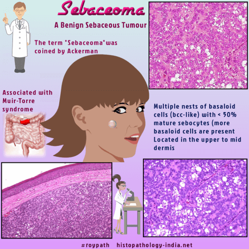
Dermpath-India

Pathology of Sebaceoma

Dr Sampurna Roy MD                        2022


Dermatopathology Quiz Case: 228

Answer -  Sebaceoma


Troy and Ackerman 
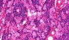
defined the term sebaceoma as benign neoplasm of basaloid cells with varying numbers of mature sebocytes. (Sebaceoma. A distinctive benign neoplasm of adnexal epithelium differentiating toward sebaceous cells. Am J Dermatopathol. 1984 Feb;6(1):7-13  )

Distinction between sebaceous adenoma and sebaceoma may be difficult and there is an increasing tendency to regard these two tumours as part of a continuum of benign tumours.

Some authors use the term sebaceous adenoma when half or less than 50% of the lesion is composed of germinative and transitional cells, and sebaceoma when greater than 50% of the lesion is composed of germinative and transitional cells.          

The term 'sebaceous epithelioma' has been largely discarded by many pathologists as the term 'epithelioma' is confusing & has been used in different ways by various pathologists.

Clinical presentation:  Presents as a solitary circumscribed nodule or an ill-defined plaque. May also present as multiple lesions, specially in Muir-Torre Syndrome.

Site: Located on the face or scalp.


Microscopic features:

Histologically, sebaceoma shows irregular shaped cell masses in which more than 50 percent cells are undifferentiated, basaloid cells together with significant aggregates of sebaceous cells and transitional cells.

Cysts and duct-like structures contain holocrine secretion and debris.

Variants:

Visit : Pathology of Rippled-Pattern Sebaceoma:

In a rare variant of sebaceoma the tumour displayed reticulated and cribriform basaloid epithelial islands.

Some cases show areas resembling seborrheic keratosis.


Differential Diagnosis:  

1) Sebaceous Carcinoma- some cases of sebaceoma are difficult to differentiate reliably from carcinoma because of the germinative cells (mitotically active and may display atypical nuclear features) ;

2) Basal cell carcinoma with sebaceous differentiation.

The tumour usually does not recur after excision.


Further reading:

A case of sebaceoma with extensive apocrine differentiation.

Sebaceoma, trichoblastoma and syringocystadenoma papilliferum arising within a nevus sebaceous.

Sebaceoma of the eyelid.

Sebaceoma of the auricle.

Immunohistochemical findings of sebaceous carcinoma and sebaceoma: retrieval of cytokeratin expression by a panel of anti-cytokeratin monoclonal antibodies.

Sebaceoma of the external ear canal: an unusual location. Case report and review of the literature.

Sebaceous lesions and their associated syndromes: part I.

Unusual malignant transformation of recurrent sebaceoma. A case report.

Characteristic Ber-EP4 and EMA expression in sebaceoma is immunohistochemically distinct from basal cell carcinoma.

Sebaceoma and related neoplasms with sebaceous differentiation: a clinicopathologic study of 30 cases.

Sebaceoma arising in association with seborrheic keratosis.  

Sebaceous carcinoma, trichoblastoma, and sebaceoma with features of trichoblastoma in nevus sebaceus.

Trichoblastoma and sebaceoma in nevus sebaceus.

Sebaceous neoplasm with reticulated and cribriform features: a rare variant of sebaceoma.


Visit:- Infectious Disease Online

Dr Sampurna Roy  MD

Consultant  Histopathologist (Kolkata - India)


Copyright © 2002-2022  histopathology-india.net