Custom Search


Dermpath-India

Pathology of Sebaceous Hyperplasia

Dr Sampurna Roy MD             2022


Dermatopathology Quiz Case: 229

Answer - Sebaceous Hyperplasia


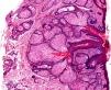
Sebaceous hyperplasia is a common benign lesion of the sebaceous gland.

This lesion has been seen in heart transplant and bone marrow recipients and is probably due to the effects of Cyclosporine.

Clinical presentation:

Solitary or multiple yellowish, umbilicated papules (1 to 3 mm in diameter).

Usually occurs in middle aged patients, but cases have been reported in younger individuals.

Juxtaclavicular beaded line - variant of sebaceous hyperplasia characterized by papules arranged in parallel rows resembling 'strands of beads'.

Site:  Face, forehead, chest, areola, mouth, vulva etc.


Microscopic features:

Histology shows separate hyperplastic sebaceous glands ; mature sebaceous lobules connect to a central dilated duct ; essentially normal cytological appearance.

Lobules consist of : 

(i) Basaloid or cuboidal cells without lipid droplets. 

(ii) Layers of mature sebocytes with abundant lipid and scalloped nuclei.


Differential diagnosis:

Nevus sebaceus (lobules are aberrant, ducts open into the epidermis, there is epidermal hyperplasia and in some cases apocrine glands)

Rhinophyma (sebaceous glands are not as well defined and grouped as in sebaceous hyperplasia).


Further reading:

Sebaceous hyperplasia within epidermis after scald.

Sebaceous gland hyperplasia of the caruncle: an uncommon diagnosis.

[Multiple sebaceous hyperplasia secondary to ciclosporin in a heart transplant patient].

Sebaceous hyperplasia of the scrotum and penile shaft.

Areolar sebaceous hyperplasia with underlying primary duct carcinoma of the breast in a woman with Donohue syndrome (leprechaunism).

Sebaceous hyperplasia: a pilot study to correlate this skin disease with circulating androgen levels.

Is "premature sebaceous hyperplasia" really a sebaceous hamartoma? Report of a case with neonatal onset.

Premature sebaceous hyperplasia in an adolescent boy.

Vulvar sebaceous hyperplasia associated with lymphedema of external genitalia.

Multiple clustered dermatofibroma with overlying sebaceous hyperplasia.

Eruptive sebaceous hyperplasia in association with chronic renal failure.

Linear sebaceous hyperplasia on the chest.

Comparative clinicopathological study of intraoral sebaceous hyperplasia and sebaceous adenoma.


Visit:- Infectious Disease Online

Dr Sampurna Roy  MD

Consultant  Histopathologist (Kolkata - India)


Copyright © 2002-2022  histopathology-india.net