

Custom Search
|


|
Dermpath-India Pathology of Nevus Sebaceus of Jadassohn
|
|
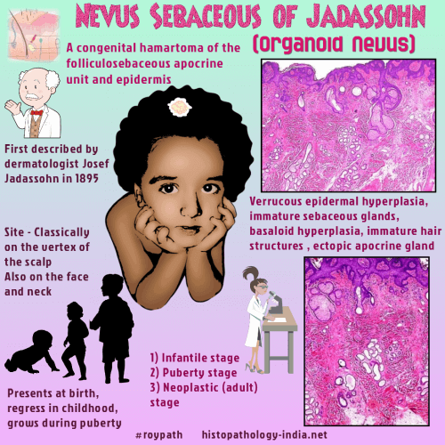
Syn: Naevus
Sebaceus ; Organoid Naevus Nevus sebaceus of Jadassohn (NSJ) is a benign, congenital hamartoma of the folliculo-sebaceous apocrine unit and epidermis. It often presents at birth, appears to regress in childhood, and grows during puberty, suggesting possible hormonal control. Nevus sebaceus of Jadassohn evolve through 3 stages. (Infantile ; Puberty ; Neoplastic in adulthood). This was first described in 1965 by Mehregan and Pinkus. If associated with features such as mental retardation, central nervous system abnormalities, oculocardiac defects, or skeletal abnormalities, it is called linear nevus sebaceous syndrome (a neurocutaneous phakomatosis). Clinical presentation: In puberty hormonal changes cause proliferation and hyperplasia of the lesion. The lesion becomes verrucous with yellow lobular structures. The lesion becomes larger, verrucous and nodular. The final stage is characterized by the appearance of nodules or tumors, with the presence of thin telangiectasias in lesions of longer evolution. Later in life, the lesions can develop benign or malignant appendageal tumors that cause further disfigurement. Site:
Located on the scalp, face
or neck as a solitary lesion and usually present since birth. Basal cell carcinoma has been observed in about 5 percent cases.
Differential diagnosis: Sebaceous hyperplasia ; Seborrheic keratosis. It may be difficult to differentiate between basal cell carcinoma and basaloid proliferation that arise in malformed hair germs in nevus sebaceous.
|
|
|
