Skin Appendage Tmour


http://www.histopathology-india.net/SAClass.htm

                   HISTOPATHOLOGY INDIA.COM

                  Microcystic Adnexal Carcinoma

         Dr Sampurna Roy MD

 
Web www.histopathology-india.net
     DermPath-India

     Site created by

 Dr Sampurna Roy MD


http://www.histopathology-india.net/dermpath.htm

March 2007

Benign sweat gland tumours

Cysts:

Hydrocystoma -Eccrine/Apocrine

Hamartomas:

Eccrine/Apocrine naevus

Eccrine Angiomatous Hamartoma

Porokeratotic eccrine ostial naevus

Benign Neoplasms:

Syringocystadenoma Papilliferum

Hidradenoma Papilliferum

Nipple Adenoma

Syringoma

Spiradenoma

Cylindroma

Chondroid Syringoma (benign mixed tumour)

Poroma

Hidradenoma

Apocrine adenoma

Papillary eccrine adenoma

Syringofibroadenoma

Malignant sweat gland tumour 

Low grade:

Microcystic adnexal carcinoma

Eccrine epithelioma

Adenoid cystic carcinoma

Mucinous carcinoma

Primary extramammary Paget’s disease

Intermediate grade:

Porocarcinoma

Digital papillary adenocarcinoma

High grade:

Hidradenocarcinoma

Apocrine adenocarcinoma

Eccrine ductal carcinoma

Spiradenocarcinoma

Malignant cylindroma

Gross examination of the skin specimen

Reporting of biopsies taken for Inflammatory Skin Diseases

Lichen planus-like lesions

Lichen Nitidus

Bullous Pemphigoid

Hailey-Hailey Disease 

Granuloma Annulare

Necrobiosis Lipoidica

Rheumatoid Nodule

Foreign body granuloma


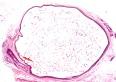
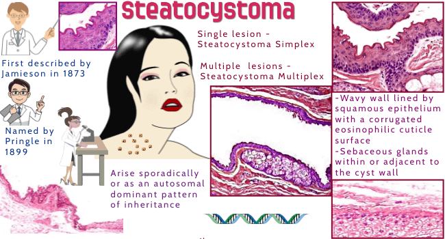
Steatocystoma presents as a solitary lesion (steatocystoma simplex) or multiple lesions  (steatocystoma multiplex) in adolescence or young adults.  

Steatocystoma multiplex is a rare benign autosomal dominant disorder of the pilosebaceous unit.  Mutations in keratin 17 have been found in some cases.

Site:   These lesions can appear virtually anywhere on the body but are more common in areas where the pilo-sebaceous apparatus is well developed, such as the trunk (especially the presternal area), neck, axilla, inguinal region, scalp, and proximal extremities.

Gross:
Macroscopically these are small, smooth, fleshy, cystic nodule.
    DermAtlas: ;

Microscopic features:    ;  

Microscopically the lesion is located in the mid dermis . The cyst has a wavy wall, lined by squamous epithelium with a corrugated eosinophilic cuticle surface. No granular layer is usually seen. Flattened lobules of sebaceous glands are usually located in the wall. The cystic space contains, keratin, vellous hair, sebum etc.Each cyst is attached to overlying normal epidermis by thin strands of undifferentiated epithelial cells.

Differential diagnosis :  Eruptive vellous hair cyst, dermoid cyst, cystic sebaceous hyperplasia.


Web www.histopathology-india.net

Abstracts:

Localized forms of steatocystoma multiplex: Case report and review of the literature.Dermatology Online Journal 2005;11(1): 22

Steatocystoma multiplex.Dermatol Online J. 2003;9(4):18.

Clinical and histologic features of 64 cases of steatocystoma multiplex.J Dermatol. 2002;29(3):152-6.

Late onset vulvar steatocystoma multiplex. Clin Exp Dermatol. 2002;27(6):445-7.

Acral steatocystoma multiplex.J Am Acad Dermatol. 2000 Aug;43(2 Pt 2):396-9

Congenital linear steatocystoma multiplex of the nose. Pediatr Dermatol. 2000;17(2):136-8

Persistent milia, steatocystoma multiplex and eruptive vellus hair cysts: variable expression of multiple pilosebaceous cysts within an affected family.Dermatology. 1998;196(4):392-6.

Steatocystoma multiplex: a case with unusual clinical and histological manifestation.Am J Dermatopathol. 1997 ;19(1):89-92.

Eruptive vellus hair cyst and steatocystoma multiplex: two related conditions?J Cutan Pathol. 1988;15(1):40-2.

Steatocystoma multiplex. A facial papular variant.Arch Dermatol. 1986 Feb;122(2):205-7.

Steatocystoma multiplex: anatomic reevaluation, electron microscopy, and autoradiography.Arch Dermatol Res. 1982;272(3-4):363-80.

Steatocystoma simplex. A solitary steatocystoma.Arch Dermatol. 1982 Jun;118(6):409-11.

Your Banner
Sebaceous tumours: Fordyce's Spots ; Steatocystoma ; Nevus Sebaceous ;Folliculosebaceous Cystic Hamartoma; Sebaceous Hyperplasia  ; Sebaceoma ; Sebaceous Adenoma;Sebaceous Carcinoma;
Soft Tissue Pathology:

Myxoid Tumours of Soft Tissue Classification of Soft Tissue Tumour;  Gross examination of soft tissue specimen ;  A practical approach to histopathological reporting of soft tissue tumours Grading of soft tissue tumours ; Lipomatous tumours ;Neural tumours ; Myogenic tumours ;Vascular tumours Fibroblastic/Myofibroblastic tumours ; Myofibroblastic tumours ;  Fibrohistiocytic tumours ; ChondroOsseous tumours ; Soft TissueTumours of Uncertain Differentiation ; Notochordal Tumour -Chordoma ;Extra-adrenal Paraganglioma ; Gastrointestinal Stromal Tumour ;

Surgical-Pathology.com;

Eye Pathology Online;

Cardiac Path Online;

Pulmonary Pathology Online;

Pathology Quiz Online;

Dermpath-India;

GI Path Online;

Mesothelioma-Online;

Soft Tissue Pathology;

Clear cell carcinoma of the skin ;

Skin Tumour;

Infectious Disease Online;

INDEX: A-D ; INDEX: E-L ; INDEX: M-P INDEX: Q-Z ; FUNGAL DISEASE ; VIRAL DISEASE.

Adnexal Tumours of the eyelid


Tumours of the Hair Follicle

Hair Germ Differentiation:

Trichoepithelioma 

Desmoplastic Trichoepithelioma

Trichofolliculoma

Trichoblastoma

Cutaneous lymphadenoma


Infundibular differentiation:

Trichoadenoma

Dilated Pore of Winer

Pilar Sheath Acanthoma

Tumour of Follicular Infundibulum

Outer root sheath differentiation:
                    
Trichilemmoma

Trichilemmal Carcinoma

Proliferating Trichilemmal Cyst (Pilar Tumour)

Matrical differentiation:
                  
Pilomatrixoma and Pilomatrix Carcinoma