Custom Search

Dermpath-India

Pathology of Trichofolliculoma

Dr Sampurna Roy MD                   2022


Dermatopathology Quiz Case: 217

Answer -  Trichofolliculoma


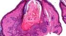
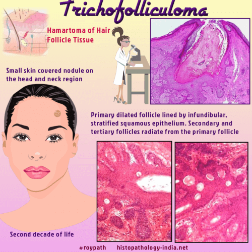
Trichofolliculoma is a hamartoma of hair follicle tissue. 

Clinical presentation:  

Presents as a small skin covered nodule on the head and neck region, usually in the second decade of life.

Tufts of vellus hair may protrude from a central umbilication.

Microscopic features: 

Key feature: Dilated infundibulocystic structure from which numerous vellus hair follicles radiate.

The tumour consists of a dilated primary follicle lined by infundibular, stratified squamous epithelium, which may be connected to the epidermis.

The lining epithelium has prominent granular layer.

Secondary and tertiary follicles are present from the central follicle.

The hair follicles are present at different levels of differentiation with numerous vellus hairs within hair follicle lumen.

Sebaceous glands may or may not be present.

Lymphocytic or granulomatous inflammation may be present in the connective tissue stroma.


Sebaceous trichofolliculoma is a variant of trichofolliculoma demonstrating numerous well differentiated sebaceous lobules emptying into the central primary follicle.

This is very closely related to folliculosebaceous cystic hamartoma.

Trichofolliculoma has an excellent prognosis.


Further reading:

Tellechea O, Cardoso JC, Reis JP, et al. Benign follicular tumors. Anais Brasileiros de Dermatologia. 2015;90(6):780-798.

A revaluation of trichofolliculoma: the histopathological and immunohistochemical features.

Inhibition of BMP signaling in P-Cadherin positive hair progenitor cells leads to trichofolliculoma-like hair follicle neoplasias.

Multiple trichofolliculomas on unusual sites: a case report and review of the literature.

Nevus lipomatosus cutaneous superficialis associated with trichofolliculoma on the lower leg.

Trichofolliculoma: a rare variant of hair follicle hamartoma.

Giant, congenital folliculosebaceous cystic hamartoma: a case against a pathogenetic relationship with trichofolliculoma.

Superficial angiomyxoma with trichofolliculoma.

Trichofolliculoma with incidental focal acantholytic dyskeratosis.  

Trichofolliculoma of the vulva associated with vulvar intraepithelial neoplasia: report of three cases and review of the literature.


Visit:- Infectious Disease Online

Dr Sampurna Roy  MD

Consultant  Histopathologist (Kolkata - India)


Copyright © 2002-2022  histopathology-india.net