Skin Appendage Tmour


http://www.histopathology-india.net/SAClass.htm

                         HISTOPATHOLOGY INDIA.COM

     Microcystic Adnexal Carcinoma

        Dr  Sampurna Roy  MD

 
 Skin Appendage Tmour


http://www.histopathology-india.net/HFoTu.htm

December 2009

Adnexal Tumours of the eyelid
Benign sweat gland tumours

Cysts:

Hydrocystoma -Eccrine/Apocrine

Hamartomas:

Eccrine/Apocrine naevus

Eccrine Angiomatous Hamartoma

Porokeratotic eccrine ostial naevus

Benign Neoplasms:

Syringocystadenoma Papilliferum

Hidradenoma Papilliferum

Nipple Adenoma

Syringoma

Spiradenoma

Cylindroma

Chondroid Syringoma (benign mixed tumour)

Poroma

Hidradenoma

Apocrine adenoma

Papillary eccrine adenoma

Syringofibroadenoma

Malignant sweat gland tumour 

Low grade:

Microcystic adnexal carcinoma

Eccrine epithelioma

Adenoid cystic carcinoma

Mucinous carcinoma

Primary extramammary Paget’s disease

Intermediate grade:

Porocarcinoma

Digital papillary adenocarcinoma

High grade:

Hidradenocarcinoma

Apocrine adenocarcinoma

Eccrine ductal carcinoma

Spiradenocarcinoma

Malignant cylindroma

Lichen planus-like lesions

Lichen Nitidus

Bullous Pemphigoid

Hailey-Hailey Disease 

Granulomatous Reaction Pattern of the Skin

Granuloma Annulare

Necrobiosis Lipoidica

Necrobiotic Xanthogranuloma

Rheumatoid Nodule

Cutaneous Sarcoidosis

Melkersson Rosenthal Syndrome

Skin lesion in Crohn's Disease

Foreign body granuloma

Rosacea

Erythema Nodosum

Folliculitis

Impetigo

Furuncle(Boil)

Carbuncle

Toxic Shock Syndrome

Sebaceous Adenoma

Sebaceous Carcinoma


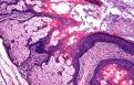
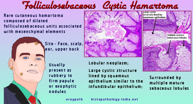
Folliculosebaceous cystic hamartoma (FSCH),  first described by Kimura et al in 1991, is a rare cutaneous hamartoma composed of dilated folliculosebaceous units associated with mesenchymal elements. Some authors have presented evidence suggesting that this lesion is very closely related to trichofolliculoma.

Clinical presentation:    Majority of lesions present as 0.5-1.5-cm papules or exophytic nodules. Lesions are usually rubbery to firm in consistency. Typically arise in adulthood but the giant variant appears to be congenital and enlarges during puberty.

Site:  Face or scalp, ear and upper back. 

Microscopic features:

Image Link

Lobular neoplasm ; large cystic structure lined by squamous epithelium similar to the infundibular epithelium ;  surrounded by multiple mature sebaceous lobules ;  rudimentary hair follicules may be present in the middle of the dermis ; the folliculosebaceous cystic structures are set in a fibrovascular stroma containing fat cells & neural tissue ; marked increase in reticular dermal collagen bundles and between sebaceous follicles ;  some areas resemble angiofibroma ; moderate perifollicular lymphocytic infiltrate.

Differential diagnosis:   

Sebaceous gland hyperplasia Steatocystoma ; Nevus sebaceus of Jadassohn  ; Small aberrant sebaceous glands, budding in close association with a hyperplastic papillomatous epidermis ;  Sebaceous trichofolliculoma- Histopathologically , FSCH shares several similar features to sebaceous trichofolliculoma, but it is usually possible to differentiate these two tumours  ; Dermoid cyst of skin.


 Abstract:

Two cases of folliculosebaceous cystic hamartoma.Clin Exp Dermatol. 2006;31(1):68-70.

Folliculosebaceous cystic hamartoma arising within a port-wine stain. Clin Exp Dermatol. 2005;30(5):509-11.

Folliculosebaceous cystic hamartoma with hair shaft fragments.Eur J Dermatol. 2005;15(2):105-6

Giant nevus lipomatosus superficialis with multiple folliculosebaceous cystic hamartomas and dermoid cysts. J Eur Acad Dermatol Venereol. 2005;19(1):84-6.

Giant folliculosebaceous cystic hamartoma of the upper extremity. J Cutan Pathol. 2004;31(3):287-90.

Genital variant of folliculosebaceous cystic hamartoma. Dermatology. 1998;197(3):258-60.

Folliculosebaceous cystic hamartoma.Pathology. 1998;30(2):212-4.

Folliculosebaceous cystic hamartoma with a neural component: an immunohistochemical study. J Dermatol. 1997;24(7):451-7.

Folliculosebaceous cystic hamartoma: a clinical pathologic study. J Am Acad Dermatol. 1996;34(1):77-81.

Folliculosebaceous cystic hamartoma with perifollicular mucinosis.
Am J Dermatopathol. 1996;18(1):58-62.

Giant folliculosebaceous cystic hamartoma. J Cutan Pathol 1994; 21 : 170-2.

Folliculosebaceous cystic hamartoma. Reported case with a neural component. Am J Dermatopathol 1993;15:277-9.

Folliculosebaceous cystic hamartoma associated with rosacea. J Dermatol. 1993;20(11):712-4.

Folliculosebaceous cystic hamartoma. A distinctive malformation of the skin. Am J Dermatopathol 1991;13:213-20.


Your Banner

Soft Tissue Pathology;

Myxoid Tumours of Soft TissueClassification of Soft Tissue Tumour;  Gross examination of soft tissue specimen ;  A practical approach to histopathological reporting of soft tissue tumours Grading of soft tissue tumours ; Lipomatous tumours ;Neural tumours ; Myogenic tumours ;Vascular tumours Fibroblastic/Myofibroblastic tumours ; Myofibroblastic tumours ; Fibrohistiocytic tumours ; ChondroOsseous tumours ; Soft TissueTumours of Uncertain Differentiation ; Notochordal Tumour -Chordoma ;Extra-adrenal Paraganglioma ; Gastrointestinal Stromal Tumour ;

Surgical-Pathology.com

Histopathology-India.net

Dermatopathology Cases

diagnostichistopathology. blogspot.com

Pathopedia-India.com

Pathology-India.com

Pancreatic Pathology Online

Gall Bladder Pathology Online

Paediatric Pathology Online

Paraganglioma-Online

Endocrine Pathology Online

Eye Pathology Online

Ear Pathology Online

Cardiac Path Online

Lung Tumour-Online

Mesothelioma-Online

Pulmonary Pathology Online

Nutritional Pathology Online

Environmental Pathology Online

Pathology Quiz Online

Dermpath-India

GI Path Online

Soft Tissue Pathology

Case Index

Infectious Disease Online; INDEX: A-D ; INDEX: E-L ; INDEX: M-P INDEX: Q-Z ; FUNGAL DISEASE ; VIRAL DISEASE.

E-book - History of  Medicine with special reference to India

Basic Pathology Blog

Tumours of the Hair Follicle

Hair Germ Differentiation:

Trichoepithelioma 

Desmoplastic Trichoepithelioma

Trichofolliculoma

Trichoblastoma

Cutaneous lymphadenoma


Infundibular differentiation:

Trichoadenoma

Dilated Pore of Winer

Pilar Sheath Acanthoma

Tumour of Follicular Infundibulum

Outer root sheath differentiation:
                    
Trichilemmoma

Trichilemmal Carcinoma

Proliferating Trichilemmal Cyst (Pilar Tumour)

Matrical differentiation:
                  
Pilomatrixoma and Pilomatrix Carcinoma

Sebaceous tumours

Fordyce's Spots

Steatocystoma

Nevus Sebaceous

Folliculosebaceous Cystic Hamartoma

Sebaceous Hyperplasia

Sebaceoma


                                                             Disclaimer  Privacy Policy  ; Advertising Policy  ;  E-mail

                                                                         Copyright © 2010  histopathology-india.net
                                                                                             All rights reserved