

Custom Search
|


|
Dermpath-India Pathology of Angiosarcoma Dr Sampurna Roy MD 2022
|

 |
|
Angiosarcomas are rare malignant mesenchymal tumours, characterized
morphologically by anastomosing vascular channels lined by atypical
and proliferative active endothelial cells.
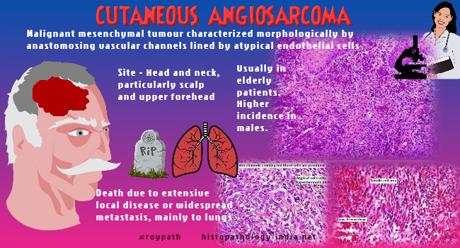
The tumour may vary from highly differentiated tumours that resemble hemangiomas to poorly differentiated anaplastic lesions that may be difficult to distinguish from carcinomas or melanomas. Angiosarcoma has a high rate of metastasis involving lungs (most commonly), liver, regional lymph nodes, bone, and other sites. Site: Angiosarcomas have a predilection for skin and superficial soft tissue (Note: Most soft tissue sarcomas are deep seated ). The sites include skin (with or without lymphedema), soft tissue, breast, liver, bone, spleen, heart and great vessels, orbit, pharynx, oral cavity, salivary gland, gastrointestinal tract, larynx, nasal cavity, kidney, prostate etc. Clinicopathological types: 1. Idiopathic angiosarcoma of the face, neck and scalp ; 2. Lymphedema associated angiosarcoma ; 3. Post-irradiation angiosarcoma ; 4. Soft tissue angiosarcoma ; 5. Angiosarcoma of the breast. Cutaneous angiosarcoma is the most common form of angiosarcoma: Age: Usually in elderly patients. Higher incidence in males. Site: Head and neck particularly scalp and upper forehead. Clinical presentation: Multifocal bruise-like areas with indurated border. Large advanced lesions are elevated, nodular and occasionally ulcerated. Grossly, the tumour extensively involves the dermis. Poorly-differentiated lesions may invade the deep structures such as subcutis and fascia. Prognosis is poor. Death is due to extensive local disease or wide spread metastasis (mainly to the lungs). Prognostic factors: External diameter of the tumour (>5 cm), depth of invasion (>3 mm), mitotic rate (>3 HPF), positive surgical margins, tumour recurrence, and metastases correlated with adverse outcome. Chronic lymphedema is the most widely recognized predisposing factor in angiosarcoma of skin and soft tissue: Lymphedema associated angiosarcomas occur in woman who have undergone radical mastectomy with removal of axillary lymphnode for breast carcinoma and have suffered from chronic severe lymphedema for many years. (Stewart Treves Syndrome) . Site: Arms in patients with lymphedema following mastectomy ; Abdominal wall following lymph node dissection for carcinoma of penis ; Arms and legs of patients with congenital lymphedema, iatrogenic lymphedema , lymphatic malformation and filarial lymphedema. Age: Elderly patients (seventh decade) in cases of lymphedema following mastectomy. Affected patients are younger (fourth or fifth decade) in cases of congenital or idiopathic lymphedema. Clinical presentation: Bluish plaque, nodules and vesicles. Post irradiation angiosarcoma presents many years after radiation therapy for benign or malignant conditions: Post irradiation angiosarcomas have occurred following radiotherapy for another malignancy such as carcinoma of the breast, cervix, ovary, endometrium and Hodgkin's disease. Cutaneous postradiation angiosarcoma of the breast (CPRASB) may occur in patients who have undergone breast-conserving therapy combined with adjuvant radiation. CPRASB differs from Stewart-Treves angiosarcoma by its shorter latency period and lack of association with lymphedema. Site: Chest wall, abdomen, shoulder, groin, flank, axilla and lower leg. Clinical presentation: The characteristic purple nodules and discoloration of the irradiated skin is the hallmark to suspect the diagnosis. Presentation ranges from small erythematous/violaceous papules or nodules to large plaques. Prognosis is poor like other types of angiosarcoma. Soft Tissue Angiosarcomas : Age: The tumour may occur at any age. These are mostly common in elderly patients. Site: Extremities or abdominal cavity (including retroperitoneum). Clinical presentation: Presents as large hemorrhagic mass. In most cases the tumour shows epithelioid cytomorphology. Miscellaneous angiosarcomas: Rare cases of angiosarcoma have been reported to arise in the setting of retained foreign material such a shrapnel, surgical sponges and adjacent to a Dacron vascular prosthesis. Angiosarcomas have occured in pre-existing benign vascular tumours including lymphangioma, 'port wine' stains and in benign and malignant peripheral nerve sheath tumours. Angiosarcomas have been reported as a complication of renal transplantation, arteriovenous fistulae, varicose ulceration, xeroderma pigmentosum and gouty tophus. There is relatively strong evidence linking various substances to the induction of hepatic angiosarcoma. About one fourth of hepatic angiosarcoma occur in patients who have received thorium dioxide (Thorotrast) for cerebral angiography, in vineyard workers exposed to AsO3-containing insectides, or in industrial workers exposed to vinyl chloride used in the production of synthetic rubber. |
|
Microscopic features:
Poorly
circumscribed dermal tumour which may infiltrate into the subcutaneous fat
and even skeletal muscle and periosteoum. - Angiomatous pattern is characterised by irregular anastomosing vascular channels dissecting through the collagen. The vessels are lined by endothelial cells with features ranging from normal appearing endothelial cells to pleomorphic, hyperchromatic cells exhibiting multilayering. Papillary processes may be present within the lumen. Numerous normal or
abnormal mitotic figures are present. - In the solid areas the tumour cell may be spindle or polygonal shaped. Vascular architecture may not be identified in the poorly differentiated areas. Reticulin stains highlights the vascular channels. - Presence of 'capillary lobule' is an indicator of angiosarcoma, in the biopsy of a patient who has received radiation therapy for breast carcinoma. - Verrucous angiosarcoma : This variant is characterized by verrucous changes with marked pseudocarcinomatous epithelial hyperplasia with hypergranulosis, compact orthokeratosis, and papillomatosis overlying an angiosarcoma in the dermis. Immunohistochemistry: CD31, CD34, and FLI1 protein appear to be the best immunohistochemistry markers of angiosarcoma; CD31 is a very useful marker in the diagnosis of angiosarcoma. CD31 is relatively sensitive and specific compared to other markers. Angiosarcoma shows membranous expression of CD31 in >90% of cases and nuclear expression of ERG, a member of the Ets family of transcription factors, in up to 100% of cases. CD34 expression is found in 50% of cases. FLI1 positivity is found in up to 100% of angiosarcomas, but its diagnostic utility is limited by a low specificity for vascular lesions. Differential diagnosis: Well differentiated angiomatous areas in angiosarcoma display cytological atypia, multilayering , papillary structures and irregular anastomosing blood vessels. Poorly differentiated angiosarcoma may resemble spindle cell sarcoma, melanoma and carcinoma. Immunohistochemical markers for keratin and S100 protein help in establishing the diagnosis. In Kaposi's sarcoma cytological atypia is less prominent and there is no endothelial multilayering. In Kaposi-like hemangioendothelioma there is lack of cytological atypia and absence of infiltrative anastomosing channels. Atypical post-radiation vascular lesions (AVLs) has been described recently. These lesions have a benign course. The time interval from radiation is significantly shorter for the development of AVL compared to angiosarcoma. The lesions are fairly well-circumscribed and confined within superficial to mid dermis and composed of complex anastomosing and focally dilated vascular spaces. Some of the vascular spaces are lined by prominent hyperchromatic endothelial cells, while others are characterized by areas with a dissecting growth pattern within dermal collagen. Endothelial multilayering is absent. |
|
Pathology of Epithelioid Angiosarcoma
|
| Angiosarcoma of the Breast: This is the most common pure malignant stromal tumour of the breast. Age: Peak incidence is in the 30s. Clinical presentation: Diffuse enlargement of breast. Gross: Tumour measures between 2 to 5 cm in diameter. Low grade tumour : Vascular, homogeneous, spongy mass with hemangioma-like cut surface. High grade tumour : Solid central area with foci of hemorrhage and necrosis. Microscopic features: Most important feature is the presence of low grade microscopic features resembling hemangioma. Prognosis depends on histologic grading: Grade1: Anastomosing vascular channels ; Tufting present. No papillary structures ; No solid anaplastic or spindle cell areas. Grade2: Endothelial tufting is present ; There are papillary structures ; Solid spindle cell areas are present.; Mitotic figures are present Grade3: Solid tumour with areas of hemorrhage, necrosis and pleomorphism; Mitotic figures are prominent. Differential diagnosis: Hemangioma ; Angiolipoma (sharply demarcated lesion, microthrombi are present)
|
|
Hemangioma -Less than 2cm in size. -Non anastomosing vascular channels lined by flattened flat non- proliferating endothelium -Non neoplastic vessels are present close to benign hemangioma -Smooth muscle fibres are identified around vascular channels
|
Angiosarcoma - Larger than 2 cm - Anastomosing, intercommunicating channels lined by 1 or 2 layers of endothelial cells. There is tufting of endothelial cells. - -
|
Angiosarcoma of the Gastrointestinal Tract : Angiosarcoma of the gastrointestinal tract is quite rare. These tumours have been reported in the stomach, small intestine, rectum, esophagus, and in the appendix. The majority of the reported gastrointestinal angiosarcomas followed previous radiation therapy, given either for malignancy (most commonly carcinoma of the cervix or ovary) or for benign lesions, such as eczema or lymphangioma. Presenting symptoms include intestinal bleeding, anemia and pain. The lesion presents either as a primary or metastatic tumour . Histologically, the tumour is characterized by a network of anastomosing, delicate vascular channels lined by atypical endothelial cells. In many cases there are sheets of malignant appearing epithelioid cells with subtle areas forming cleft-like spaces suggestive of vascular differentiation. The typical immunohistologic pattern is positivity for vimentin and endothelial markers (eg, factor VIII–related antigen, UEA-I, CD31, and CD34), The large, plump, polygonal cells in epithelioid angiosarcoma may be diffusely and strongly positive for cytokeratins and only show subtle signs of vascular differentiation, creating potential diagnostic confusion with primary or metastatic carcinoma, melanoma and mesothelioma, as well as sarcomas with epithelioid cytomorphology . Immunohistochemistry with vascular markers, CK20, and S-100 protein may be helpful in differentiating angiosarcoma from carcinoma and melanoma. Given the clinically aggressive behavior of angiosarcoma, proper classification and treatment is important. Cardiac Angiosarcoma: Most common primary malignant tumour of the heart. It commonly causes blood flow abnormalities, extensively infiltrates cardiac structures, and may extend through the heart wall to involve adjacent structures. Age: This disease is most commonly found in middle-aged men. Clinical presentation: May present with congestive heart failure and dyspnea. Patients with pericardial lesions present with pericarditis. Most patients are dead within 10 months of the onset of symptoms. Site: The tumour is most often located in the right atrium. The two major forms: i) large obstructing mass ii) locally infiltrative tumour. Diagnosis is made on surgical biopsy specimen, pericardial fluid cytology and sometimes on endomyocardial biopsy. Microscopic features: The histopathology vary from well differetiated areas to poorly differentiated solid areas. Well differentiated areas are characterized by numerous vascular areas in which neoplastic cells are loosely and irregularly arranged to form incomplete vessels or anastomosing blood-filled vascular channels. Less-differentiated solid areas irregularly surround the differentiated vascular areas. Spindle- shaped anaplastic cells are present in the solid less differentiated areas. Primary cardiac Epithelioid angiosarcoma has also been reported. Differential diagnosis: Poorly differentiated sarcoma; metastatic tumour ; fibrous (sarcomatoid) mesothelioma Cardiac angiosarcoma. Histological, immunohistochemical and ultrastructural study. Cardiac angiosarcoma: a case report.
|
|
|
|
|
