

Custom Search
|

|
Infectious Disease Online Pathology of Histoplasmosis due to Histoplasma Duboisii (African Histoplasmosis)
|
|
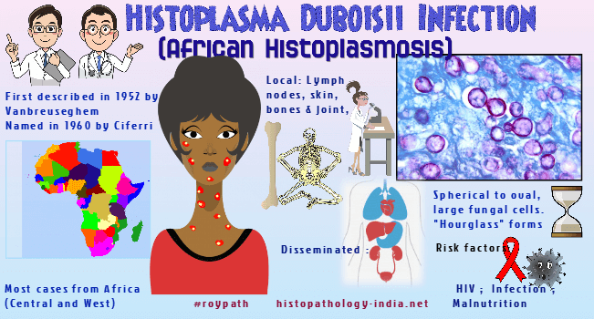
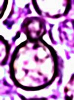
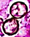
Syn: African histoplasmosis, Large histoplasmosis, Largeform histoplasmosis. Histoplasma duboisii is an invasive fungal organism which commonly affects lymph nodes, bones and skin. African histoplasmosis often arises in patients already suffering from tuberculosis, schistosomiasis, malnutrition, leishmaniasis, or AIDS. The duboisii variety of Histoplasma capsulatum was first described in 1952 (by Vanbreuseghem) named in 1960 (by Ciferri). It was fully described as a clinical disease in 1964 (by Cockshott and Lucas). The organism was obtained from soil mixed with bat guano, as well as the bowel of bats in 1991 (Gugnani et al.). It has been identified in two species of baboons from Guinea and Senegal. Histoplasmosis is a deep mycosis due to a fungus, Histoplasma capsulatum, which possesses two varieties, namely, Histoplasma capsulatum var. Capsulatum, an agent of the so-called American histoplasmosis , and Histoplasma capsulatum var. duboisii, an agent of African histoplasmosis. When grown in vitro, mycelial and yeast forms of this fungus are indistinguishable from those of the classical, small-celled form or capsulatum variety of this species. The two can be distinguished from each other only when the size of the yeastlike cells that develop in tissue is observed. Histoplasma capsulatum var. duboisii differs morphologically from H. capsulatum var. capsulatum by the typical production of a large-celled yeast form. The yeast cells average about 10 μm in diameter. They are thick walled and round or oval. Diseases caused by the two varieties of Histoplasma capsulatum are clinically and pathologically distinct. Natural infection caused by Histoplasma capsulatum var duboisii has been reported mostly in human and nonhuman primates from Africa. It is an important deep mycosis endemic in Central and West Africa and in the island of Madagascar. The disease is rarely seen in the United States in persons who previously lived or travelled in Africa.
Lesions typically contain a dispersed granulomatous inflammatory reaction in which large numbers of yeast-like cells are seen within the cytoplasm of histiocytes and huge multinucleated giant cells.
Tissue forms of H. capsulatum var. duboisii and Blastomyces dermatitidis are of similar size and shape and thus may be mistaken for each other. However, the latter buds by a broader base and is multinucleated. Histoplasmosis capsulati also occurs in Africa, but its causative agent is much smaller (2 to 4 micrometer) in tissue than the large-celled duboisii variety. Amphotericin B and excision of isolated skin lesions are treatment of choice for disseminated and localized infections, respectively.
|

|
|
