

Custom Search
|


|
Dermpath-India Pathology of Hyalinizing Spindle Cell Tumour with Giant Rosettes Dr Sampurna Roy MD 2022
|

|
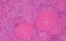
Hyalinizing spindle cell tumour with giant rosettes was first reported by Lane et al in 1997. Hyalinizing spindle cell tumor with giant rosettes: a distinctive tumor closely resembling low-grade fibromyxoid sarcoma.Am J Surg Pathol. 1997 Dec;21(12):1481-8. It is now regarded as a variant of low-grade fibromyxoid sarcoma. It is a rare low-grade sarcoma with indolent behavior, and wide resection of the tumour with prolonged follow-up are needed because patients may develop late metastases. Age: This painless, slow growing tumour can develop at any age. It commonly occurs in young and middle aged adults (average age 38 years). Site: The lesion is commonly located in the deep soft tissue of the extremities, particularly the thigh. Other sites include the chest wall, axilla, buttock and the neck. Gross features: These are deep seated, oval, multilobulated , well circumscribed, pale, firm tumours. The lesion usually ranges from 5-10 cm in maximal dimension. Focal pseudocystic degeneration may be present. Microscopic features: The tumour consists of uniform oval to spindle cells with fibroblastic or neural-like appearance. These are arranged in fascicles in a collagenous or myxoid stroma. The most striking feature is the presence of hyalinized nodules surrounded by round or ovoid tumour cells giving the appearance of rosette or palisaded granuloma. The eosinophilic area at the centre of the rosettes is composed of collagen fibrils. Pleomorphism is minimal. Mitoses are scarce. In small number of cases there may be hypercellular areas with cells showing nuclear enlargement and hyperchromatism. The miototic activity may range from 1 to 20 per 10 high-power fields. Immunohistochemistry: Spindle cells are immunoreactive for vimentin, HAM56, factor XIIIa , collagen IV, and CD68. The rounded cells comprising the collagen rosettes are positive for vimentin, S100 protein, Leu7(CD57) , synaptophysin, protein gene product (PGP) 9.5 , neuron specific enolase and sometimes CD34. The cells are negative for desmin, alpha-smooth muscle actin , keratin, and epithelial membrane antigen. Differential diagnosis: Fibromatosis ; Neuroblastoma like neurilemmoma ; Fibrosarcomatous DFSP with giant rosettes :
|
|
|
