

Custom Search
|


|
Dermpath-India Pathology of Nerve Sheath Myxoma |
| Syn:
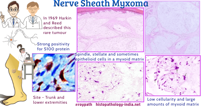
Dermal Nerve Sheath Myxoma Nerve sheath myxoma is a benign soft tissue tumour. In 1969 Harkin and Reed described an unusual myxoid tumour of probable nerve sheath origin and named it myxoma of nerve sheath. Later in 1980, Gallager and Helwig described similar lesions as Neurothekeoma. Cellular neurothekeoma was described by Barnhill and Mihm in 1990. Recent gene studies strongly supports that, nerve sheath myxomas and neurothekeoma are distinct neoplasms. Schwann-cell differentiation in the electron microscopic examination of Nerve sheath myxoma cells, suggests an origin from nerve sheath precursor cells. However, there are still some theories proposing a proliferation of other perineural cells, which brings controversies to the cell of origin. There are some similar features of Nerve sheath myxoma with other neural tissue tumors, like schwannoma and neurofibroma. That seems the reason why various names such as pacinian neurofibroma, cutaneous lobular neurofibroma and perineural neurofibroma have been used for this lesion.
Ref:
Malkoc M, Ormeci T, Keskinbora M, Yılmaz A,
Korkmaz O, Tanik CB. Nerve sheath myxoma of the dorsal paravertebral
space. International Journal of Surgery Case Reports.
2014;5(11):858-860. doi:10.1016/j.ijscr.2014.10.003. Microscopic features:
Nonencapsulated, multilobulated and hypocellular tumour, usually located in the reticular dermis and often extends into the subcutaneous fat. Tumour is composed of spindle cells, stellate and sometimes epithelioid cells set in a myxoid stroma. Spindle cells are arranged in swirling and concentric fashion. Sometimes multinucleate cells are present. Focally, there may be nuclear atypia and normal mitotic figures are often present. Rarely, foci of ossification and cartilaginous metaplasia have been reported. Immunohistochemically, the tumour is S100 protein positive. Differential Diagnosis: Myxoid Tumours of Soft Tissue : The differential diagnoses include schwannoma, neurofibroma, neurilemmoma, leiomyoma, intramuscular myxoma and low-grade sarcoma. Nerve Sheath Myxoma - Pathology Infographics
|
|
Further reading:
Neurothekeoma: report of a case in an infant and review of the literature.
|
|
|
