

Custom Search
|

|
Custom Search
|
Infectious Disease OnlinePathology of Opisthorchiasis
|
|
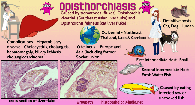
Opisthorchiasis is a parasitic disease that can infect fish eating mammals, including humans. The trematode liver fluke worms, Opisthorchis viverrini and Opisthorchis felineus, cause opisthorchiasis by parasitizing the liver and biliary passages of their hosts. |
|
Opisthorchiasis is caused by the small liver flukes
Opisthorchis viverrini and
Opisthorchis felineus. Opisthorchis felineus is the prominent fluke in Russia, Ukraine, and Kazakhstan. In the European Union, sporadic human infections have been documented in Germany, where the parasite has been detected in red foxes and cats, and in Greece. In Italy, O. felineus was first described in cats and dogs in Pisa (Tuscany Region) and in cats in Turin (Piedmont Region). Cases of human infection, were reported in 2003 and 2005, when 2 outbreaks of opisthorchiasis occurred after persons consumed fish from Lake Trasimeno (central Italy). Opisthorchis viverrini (OV) infection is a major public health problem in the Mekong River Basin region of Southeast Asia, especially in Thailand, the Lao People’s Democratic Republic (Lao PDR), Cambodia, and Vietnam. The first case of opisthorchiasis was reported in 1911 by Leiper in the post-mortem examination of two prisoners from a jail in Chiengmai, northern Thailand. Later on Sadun in 1953, Harinasuta and Vajjarasthira in 1961, and Wykoff in 1965 had demonstrated a complete life cycle of O. viverrini. Opisthorchis viverrini has a complicated life cycle with 2 intermediate hosts, a freshwater snail is the first intermediate host , and a freshwater fish is the second intermediate host. Embryonated eggs are passed the faeces which are ingested by snails (first intermediate host). After several stages of development in the snail, the cercariae are released from the snail which enter into fresh water fish (second intermediate host) and encyst in the muscle or under the scales. Cats, dogs, and various fish-eating mammals including humans are the definitive host. Humans have become infected by ingesting undercooked or raw fish containing infective metacercariae. After ingestion, the metacercariae excyst in the duodenum and ascend through the ampulla of Vater into the biliary ducts. Here they attach and develop into adults and lay eggs. [ Source of infection: Undercooked and raw fish: (1) Koi pla, eaten soon after preparation; (2) Moderately fermented pla som; stored for a few days to weeks; and, (3) Pla ra extensively fermented, highly salted fish, stored for at least 2-3 month.] Adult flukes reside in small- and medium-sized intrahepatic bile ducts, and occasionally also in the gallbladder, extrahepatic bile ducts, and in the pancreatic duct. Opisthorchiasis is associated with a number of hepatobiliary diseases, including cholangitis, obstructive jaundice, hepatomegaly, cholecystitis, biliary lithiasis and cholangiocarcinoma. Cholangiocarcinoma is highly prevalent in Asian countries, particularly in Thailand, China, Taiwan and Korea. The principal risk factor for Cholangiocarcinoma in this region is infection with the human liver flukes, Opisthorchis viverrini and Clonorchis sinensis, which are Group 1 carcinogens as classified by the World Health Organization. Three main mechanisms are proposed to contribute to Cholangiocarcinoma through chronic infection with O. viverrini: (1) mechanical damage to the biliary epithelia caused by the feeding activities of the parasites, (2) immunopathology caused by infection-related inflammation, and (3) toxic effects of parasite excretory/secretory molecules In contrast to infection with O. viverrini, many patients infected by O. felineus suffer from fever and hepatitis-like symptoms in the early stage of infection, and no association with cholangiocarcinoma formation has been established. In chronic infections, patients may present with cholangitis and liver abscess because of biliary obstruction. Diagnosis is achieved by detection of eggs in faeces, however, species-specific diagnosis on the basis of egg morphology is difficult. Polymerase chain reaction detecting DNA of the adult parasite in stool may be helpful and sometimes the parasites are detected by microscopic examination. Visit: Pathology of Clonorchiasis
|
|
|

Consultant Histopathologist (Kolkata - India)
|
![]()
Copyright © 2021 histopathology-india.net