Custom Search


Dermpath-India

Pathology of Xanthelasma

Dr Sampurna Roy MD                 

Dermatopathology Quiz Case: 194

Answer -  Xanthelasma Palpebrarum


 Syn: Xanthelasma Palpebrarum.     

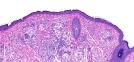
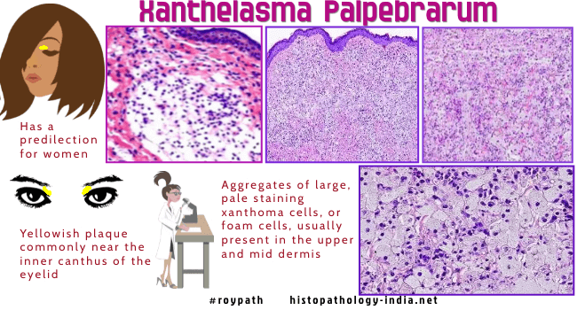
Xanthelasma is the most common form of xanthoma and is characterized by one or more yellowish plaques on the eyelid or in the periorbital skin.

Most patients are in the 5th or 6th decade of life.

The patients are often associated with atherosclerosis, and coronary artery disease.

Lipid levels are normal in about 50% of the patients.

In some young patients there is higher incidence of hypercholesterolemia.

Alteration in apolipoprotein levels (A1 and B) in patients with xanthelasma may predispose to cutaneous and systemic deposition of lipids, including atherosclerosis.

Patients irrespective of the size of the lesion or serum lipid levels should be screened using carotid intima media thickness for detection of subclinical atherosclerosis.    


Microscopic features:  

There are small aggregates of large, pale staining foam cells in the upper dermis. Inflammatory cells are not present and usually there is no fibrosis.

Xanthelasma are not always limited to the superficial dermis but  may penetrate the entire dermis and reach into the orbicularis muscle.


These lesions are usually removed for cosmetic reasons.

Depth of tissue invasion of xanthelasma plays an important role in the selection of suitable surgical procedure.

A careful clinical examination of the famous painting of Mona Lisa by Leonardo da Vinci shows a yellow irregular spot at the inner end of the left upper eyelid and a 3 cm long well-circumscribed lump on the dorsum of the right hand beneath the index finger.

According to several authors this is regarded as the first case of Xanthelasma.

Related post: Verruciform Xanthoma  

Dermatopathology Case 30

The painting Mona Lisa in the Louvre, Paris, by Leonardo da Vinci (1503-1506), shows skin alterations at the inner end of  the left upper eyelid similar to xanthelasma, and a swelling of the dorsum of the right hand suggestive of a subcutaneous lipoma.

These findings in a 25-30 year old woman, who died at the age of 37, may be indicative of essential hyperlipidemia, a strong risk factor for ischemic heart disease in middle age.

As far as is known, this portrait of Mona Lisa painted in 1506 is the first evidence that xanthelasma and lipoma were prevalent in the sixteenth century, long before the first description by Addison and Gall in 1851.

Xanthelasma and lipoma in Leonardo da Vinci's Mona Lisa. Isr Med Assoc J. 2004 Aug;6(8):505-6.

Ref: The Real Code of Leonardo da Vinci


Further reading:

Xanthelasma palpebrarum: a marker of premature atherosclerosis (risk of atherosclerosis in xanthelasma).

Xanthelasma palpebrarum treatment with a 1,450-nm-diode laser.

[The underestimated depth of tissue invasion of xanthelasma--a histological study].

Cardiovascular profile of xanthelasma palpebrarum.

Normolipemic papular xanthoma with xanthelasma.

Xanthelasma: clinical indicator of decreased levels of high-density lipoprotein cholesterol.

Xanthelasma palpebrarum and its relation to atherosclerotic risk factors and lipoprotein (a).


Visit:- Infectious Disease Online


Dr Sampurna Roy  MD

Consultant  Histopathologist (Kolkata - India)


Copyright © 2002-2022  histopathology-india.net