

Custom Search
|
|
Custom Search
|
|
Dermpath-India Pathology of Verruciform Xanthoma Dr Sampurna Roy MD 2022
|
| Verruciform xanthoma
(VX) is a rare, benign, mucocutaneous, nondestructive lesion characterized by proliferation of non-Langerhans
lipid-rich histiocytes.
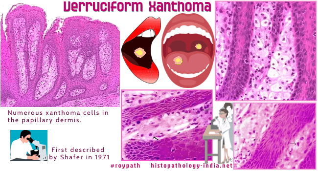
Clinically the lesion may mimic cutaneous squamous cell carcinoma and a correct diagnosis is crucial to avoid inappropriate aggressive treatment. It was first described by Shafer in 1971 where he described 15 cases of this condition in the oral cavity and coined the term 'verruciform xanthoma'. (Shafer WG. Verruciform xanthoma. Oral Surg Oral Med Oral Pathol. 1971;31:748–9) Related post: Pathology of Xanthelasma Human papillomavirus has been indicated in several cases of VX as a causative agent. Cases have been reported in immunocompromised patients with HIV-1 infection and graft versus host disease. Verruciform xanthoma may be associated with epidermal nevi, fibroepithelial polyp of the vulva, squamous cell carcinoma, arteriovenous hemangioma, discoid lupus erythematosus and lymphedema of the leg. Site: Predominantly noted in the oral cavity, but it has been reported to occur on the genital skin and mucosa (vulva, perianal skin, scrotum, penis). Gross: Usually solitary, flat plaques or warty lesions (about 2 cm in diameter). Microscopic features: Hyperkeratosis, focal parakeratosis and verrucous acanthosis. Exocytosis of neutrophils into the upper layer of epithelium and the parakeratotic scales. Characteristic feature: Presence of numerous xanthoma cells in the papillary dermis. Cells contain lipid and small amounts of PAS-positive diastase resistant material. Immunohistochemistry : The characteristic foam cells of verruciform xanthoma showed strong positive staining for CD68 [KP1] and vimentin and weak positivty for cytokeratin. S100 protein is negative. Differential diagnosis: Differential diagnosis also included squamous cell carcinoma, verrucous carcinoma, seborrheic keratosis, verruca vulgaris, condyloma acuminatum, granular cell tumor with pseudoepitheliomatous hyperplasia.
|
|
|
Visit:-
Infectious Disease Online

Consultant Histopathologist (Kolkata - India)
|
![]()
Copyright © 2022 histopathology-india.net


