Custom Search
|


|
Dermpath-India Pathology of Angiolipoma Dr Sampurna Roy MD 2023
|

| Age:
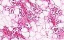
Angiolipoma
occurs in young adults
Site: These subcutaneous tumours are usually located on the forearm, arm and chest wall. Clinical presentation: Presents as multiple, painful yellow, firm, circumscribed tumours. Angiolipoma usually do not recur after excision. New lesions may continue to develop elsewhere. Microscopic features: -Thin fibrous capsule with fibrous septa dividing the lesion into lobules. -There are mature adipocytes together with groups of small vessels. - Presence of fibrinous microthrombi in the lumen is a diagnostic feature. - In 'cellular angiolipoma' the vascular component is more than 90%. - Prominent pericytes may be present around the capillaries. - Numerous mast cells are noted throughout the tumour. - Degenerative changes (i.e. hyalinization, myxoid change and fibrosis) may be present in longstanding cases. - Angiomyxolipoma is characterized by myxoid stroma. Unlike angiolipoma which has normal karyotype, angiomyxolipoma shares cytogenetic changes with lipoma, spindle cell lipoma and myxoma. Two cases of angiomyxolipoma (vascular myxolipoma) of subcutaneous tissue. Angiomyxolipoma (vascular myxolipoma) of subcutaneous tissue. Angiomyxolipoma shares cytogenetic changes with lipoma,spindle cell/ pleomorphic lipoma and myxoma. Cytogenetic: Normal karyotype Differential diagnosis: One must exclude well-differentiated angiosarcoma of the breast from angiolipomas arising on the chest wall. Cellular angiolipomas may resemble Kaposi's sarcoma or primitive capillary hemangioma. |
|
|