

Custom Search
|


|
Dermpath-India Pathology of Spindle Cell Lipoma and Pleomorphic Lipoma Dr Sampurna Roy MD 2022
|
 |
|
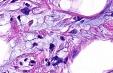
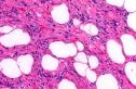
Spindle cell lipoma and pleomorphic lipoma occur in adults and are more common in males. These are located on the back of the neck, upper back or shoulders. These tumours usually present as a solitary, painless, slow growing, lobular mass with an average diameter of 5 cms. Rarely multiple lesions may develop. Cut surface has a yellow or grayish - yellow colour. Sometimes mucoid areas are present. Histological features of Spindle cell lipoma: This is a well circumscribed lesion usually confined to the subcutis. Some lesions are confined to the dermis (intradermal spindle cell lipoma). The tumour consists of an admixture of adipocytes and spindle cells. Spindle cells are arranged in short fascicles. The spindle cells are small and elongated and contain bland, uniform nuclei and pale eosinophilic cytoplasm. In areas nuclear palisading is present. The stroma is characterized by the presence of eosinophilic collagen fibres. Numerous mast cells are present. There is no increase in mitoses. Some cases show prominent myxoid changes. The adipocytes are mature. No lipoblasts are seen. CD34 is extensively positive. Histological features of Pleomorphic Lipoma : This is a well circumscribed tumour which is usually superficial in location. The tumour consists of mature adipocytes, abundant interstitial collagen, together with bizarre, often multinucleated cells (floret giant cells). There is dense collagen. Myxoid stroma contains arborizing vessels. No lipoblasts are present. (Some lipoblast - like cells may be seen) Mitoses is usually absent.
There is minimal adipocytic
nuclear atypia. Differential diagnosis of spindle cell lipoma and pleomorphic lipoma: Differential diagnosis of spindle cell lipoma- Schwannoma and neurofibroma (Adipocytes absent, S100 protein positive ). In well differentiated liposarcoma (atypical lipoma)-sclerosing variant careful clinico- pathological correlation is important. Unlike pleomorphic lipoma these are usually located in deeper tissue. There is variation in size of adipocytes together with other atypical features. Scattered lipoblasts are also present. Differential diagnosis of pleomorphic lipoma also includes pleomorphic liposarcoma.
|
|
|
