

Custom Search
|


|
Dermpath-India Pathology of Blue Nevus "A benign pigmented tumour with bluish gray appearance" Dr Sampurna Roy MD 2023
|

|
Syn: Tieche's
nevus ; Blue nevus of Jadassohn-Tieche Blue naevus was first described by Tiesche in 1906. These lesions may cause diagnostic difficulty in case of large size, involvement of subcutaneous tissue, asymmetrical pigmentation and presence of lymphnode metastasis.
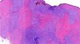
The macroscopic bluish appearance is due to the presence of deep intradermal melanin pigment viewed through intact skin (Tyndall effect). Site: Although it usually occurs in skin, it has been reported in other locations, such as oral mucosa, sclera, uterine cervix, vagina, prostate, spermatic cord, pulmonary hilus, orbit, conjunctiva, maxillary sinus, breast, and lymph nodes. The two main variants: 1) Common Blue Nevus:
1) Common Blue Nevus:
These are usually located on the head, hands and feet.
Occasionally, the lesion is hypomelanotic (Differential diagnosis: Dermatofibroma). Usual differential diagnosis include nevi of Ito, Ota and the Mongolian spot.
These acquired lesions are usually located in the sacrococcygeal/gluteal region and less common sites include face, scalp and extremities. These lesions clinically presents as nodule, tumour or plaques, usually 1 - 2 cm in diameter (sometimes much larger).
The differential diagnosis include malignant melanoma,
fibrous histiocytoma,
pigmented dermatofibrosarcoma protuberans
and
schwannoma. Other
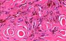
variants of Blue Nevus: Microscopically, the lesion consists of two cell types: (i) globular to fusiform and heavily pigmented (ii) polygonal or spindle shaped and light pigmented or non pigmented. Atypical Cellular Blue Nevus: Proposed by Mihm et al, the lesion demonstrates clinico-pathological features intermediate between cellular blue nevus and malignant blue nevus. There is evidence of architectural and cytological atypia together with necrosis. No atypical mitoses were present and there was no evidence of metastasis. Malignant Blue Nevus: The patient usually complains of sudden increase in size of a longstanding lesion or there may be history of repeated unsuccessful local excision. The lesion includes clear cut areas of malignant and benign blue nevus, or the histological appearances may resemble cellular blue nevus together with pleomorphism prominent atypical mitoses and areas of coagulative necrosis. Follow up of the cases reveal significant morbidity and death by metastatic disease.
Blue Nevus with Lymphnode metastasis: Blue naevus with lymphnode metastasis is characterised by cellular blue naevus in the skin associated with the finding of melanocytes within a regional lymphnode. In contrast to melanoma cells, blue nevus cells within lymphnodes are confined to capsule and do not show atypical features or mitosis. It has been suggested that the presence of cells of a blue nevus within a lymphnode is due to error during migration of melanocytes from the neural crest to the skin.
|
|
|
Visit:-
Infectious Disease Online
 Prof (Dr) Haradhan Roy MD (AIIMS) (1928-2022) (R) Director-Professor and Head of the Dept of Pathology, Calcutta National Medical College, Calcutta University India |

Consultant Histopathologist (Kolkata - India)
|
![]()
Copyright © 2002-2023 histopathology-india.net