

Custom Search
|


|
Dermpath-India Pathology of Dermatofibrosarcoma Protuberans Dr Sampurna Roy MD 2022
|

|
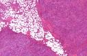
Dermatofibrosarcoma
Protuberans
(DFSP) is a
fibrohistiocytic tumour of intermediate malignancy, characterized by a
distinctive storiform growth pattern and frequent local recurrences. This tumour was first described in 1924 by Darier and Ferrand as 'progressive and recurring dermatofibroma'. Over the years this tumour has been considered to be of fibroblastic, histiocytic and even of neural origin by different authors. Inview of the similarity of the tumour with benign fibrous histiocytoma, this tumour is classified with fibrohistiocytic tumours. Dermatofibrosarcoma Protuberans usually occurs in young and middle aged adults and is commonly located in the head and neck region followed by upper extremity. Palms and soles are not affected. Cytogenetic analysis reveals reciprocal translocation t(17;22)(q22;q13) and ring chromosome derived from translocation r(17;22). Macroscopically, the tumour presents as a firm and grey white plaque, exophytic nodule or a massive pedunculated tumour. The pigmented variant has a slate-gray appearance. Microscopic features: - Dermal tumour which extends into the subcutis where it infiltrates around small groups of fat cells in a lacy or linear fashion. - The tumour is composed of interwoven bundles of spindle cells with plump nuclei arranged in a storiform or cartwheel pattern. - Superficial grenz zone is present separating the tumour from the epidermis. - The overlying epidermis is normal or atrophic. - Scattered mitotic figures are present ( not more than 5 per 10 high power fields). - The tumour cells surround the dermal appendages. The appendages are not destroyed by the tumour. - Other features include presence of thin walled blood vessels, occasional Touton giant cells, foam cells and granular cells. - Immunohistochemistry - The most diagnostic marker is CD34 (human progenitor cell antigen). 50-100% cells show positivity.
Fibrosarcomatous change in Dermatofibrosarcoma Protuberans Fibrosarcomatous change in Dermatofibrosarcoma protuberans represents a form of tumor progression in DFSP and is associated with a significantly more aggressive clinical course than in ordinary DFSP, indicating a possible need for treatment intensification in such cases. Features include increased cellularity and mitoses (more than 8 mitoses per 10 high power field). Other features include focal myxoid change, keloid like hyalinization, giant rosettes, pigmented melanocytes, myoid nodules and bundles. Less than 50% cells display CD34 positivity. The tumour may be associated with metastasis. Myxoid variant of Dermatofibrosarcoma Protuberans: Storiform pattern is less prominent. Blood vessels are more conspicuous. Features supporting myxoid DFSP - (1) CD34: Positive (2) Pattern of infiltration of the adipose tissue.
(3) Genetic alterations:
ring / markers t (17;22) Indeterminate Fibrohistiocytic Lesions: This tumour is characterized by combined features of dermatofibroma and Dermatofibrosarcoma protuberans. Features include- - Keloidal collagen - Infiltration of the subcutis in a honeycomb pattern - Low mitotic count - Dual population of CD34 and factor XIIIa positive cells Differential Diagnosis: Giant cell fibroblastoma: a report of three cases with histologic and immunohistochemical evidence of a relationship to dermatofibrosarcoma protuberans. - Dermatofibroma - Dermatofibromas are small (often less than 1 cm), symmetrical and usually do not penetrate adipose tissue. Hemosiderin pigment is present. Tumour border is usually infiltrative but regular. Collagen is present at the peripheral border, in a keloidal pattern. There is birefringence of the collagen in dermatofibroma with polarization. Unlike Dermatofibrosarcoma protuberans, adnexal structures are not entrapped within the lesion. Heterogenous population of cells (histiocytes, fibroblasts or mixed). In Dermatofibrosarcoma protuberans, the cells are usually regular, small spindle shaped. - Neurofibroma - Neurofibromatous changes in dermatofibrosarcoma protuberans: a potential pitfall in the diagnosis of a serious cutaneous soft tissue neoplasm. - Atypical Fibroxanthoma and Pleomorphic Sarcoma (Malignant Fibrous Histiocytoma) -There is prominent cellular pleomorphism in these lesions. - Classic Fibrosarcoma - Deep seated tumour. High mitotic index. The tumour rarely extends to the dermis - Fasciitis The cells display uniform ' tissue culture' growth pattern. Differential diagnosis in Myxoid Dermatofibrosarcoma Protuberans - - Myxoid Neurofibroma- Immunohistochemistry is particularly useful in this condition. - Myxoid Liposarcoma- Presence of lipoblasts. - Myxofibrosarcoma - Presence of pleomorphic cells.
|
|
Further reading: Transformed dermatofibrosarcoma protuberans: a clinicopathological study of eight cases. FNA diagnosis of dermatofibrosarcoma protuberans. Cellular digital fibromas: distinctive CD34-positive lesions that may mimic dermatofibrosarcoma protuberans. Atrophic dermatofibrosarcoma protuberans: a case report and reappraisal of the literature. Differential expression of HMGA1 and HMGA2 in dermatofibroma and dermatofibrosarcoma protuberans: potential diagnostic applications, and comparison with histologic findings, CD34, and factor XIIIa immunoreactivity. Dermatofibrosarcoma protuberans, giant cell fibroblastoma, and hybrid lesions in children: clinicopathologic comparative analysis of 28 cases with molecular data--a study from the French Federation of Cancer Centers Sarcoma Group.Am J Surg Pathol. 2003;27(1):27-39
|
|
|

![]()
Copyright © 2002-2022 histopathology-india.net