

Custom Search
|

|
Infectious Disease Online Pathology of Cystoisosporiasis (Infection caused by Cystoisospora Belli)
|
| Syn:
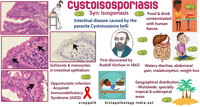
Isosporiasis Cystoisospora belli (formerly known as Isospora belli), is a protozoal parasite in the class of sporozoa, a group that includes toxoplasma and cryptosporidium. Cystoisospora belli in humans, has been described as causing chronic diarrhea and acalculous cholecystitis in patients with the acquired immunodeficiency syndrome (AIDS). Isospora belli was first discovered by Rudolf Virchow in 1860. Clinical
presentation:
Cystoisosporiasis should be suspected in HIV-infected patients from tropical
countries and the organism (Cystoisospora belli ) has been commonly identified in the small bowel of patients with
mucous diarrhoea, weight loss, fever, peripheral eosinophilia ,
malabsorption and low CD4+ cell counts. Disseminated extra-intestinal isosporiosis may sometimes occur in AIDS patient. Investigation: When oocysts cannot be identified in stool or intestinal aspirate, an upper endoscopic small biopsy can be diagnostic. Histological features: Moderate to severe enteritis, villous shortening , crypt hyperplasia, disorganised epithelium with loss of nuclear polarity, cytoplasmic vacuolation and intraepithelial lymphocytosis may be noted. Infiltration of lamina propria with eosinophils, neutrophils, lymphocytes and plasma cells may occur. Schizonts and gametocytes are identifiable within the epithelial cells. Merozoites (the asexual form) is most readily identifiable from the villous base to the tip. It forms clusters of small crescent-shaped structures to single large uninucleated banana shaped forms, similar in size to enterocyte nuclei. The merozoites are found in perinuclear and often subnuclear locations surrounded by pale parasitic cytoplasm. Special stains: The organisms can be seen on routine haematoxylin and eosin staining. Alcian blue stain highlights the parasites. Giemsa stain provides an even better contrast. Differential diagnosis: Cryptosporidium: Restricted to apical portion of the enterocyte.
|
|
|
