HISTOPATHOLOGY INDIA.COM        

    Adult Respiratory Distress Syndrome

      Dr  Sampurna Roy  MD

 
Web www.histopathology-india.net
 
  Gastrointestinal Stromal Tumour


http://www.histopathology-india.net/Infection.htm


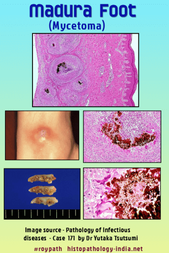
Mycetoma is a localized chronic, and deforming granulomatous infectious disease of subcutaneous tissues, skin and bones,that is present worldwide and endemic in tropical and subtropical regions.

Mycetoma is a pathological process in which the causative agents - a fungus (eumycetoma) or a bacterium (actinomycetoma) -  from exogenous source produce grains.  Distinction between eumycetoma and actinomycetoma is very important for the treatment.

Geographic distribution:  Seen in tropical region e.g. Asia, Africa, central and south America.     Vist:  Infectious Disease Online

Mode of infection: Organisms are normally present in environment (soil & dust). Infection occurs in bare-footed persons after minor penetrating skin injury inoculating soil organisms, occurring preferentially in rural areas, usually among labourers who work barefoot.

Age and sex:  Mycetoma commonly affects adults aged 20 to 40 years, predominantly males.

Site: It is a chronic, tumorous, fungal infection of deep soft tissues and bones of foot (Madura foot) . Foot is most commonly affected.

Clinical presentation : Both forms of mycetoma present as a progressive, cutaneous and subcutaneous swelling, although actinomycetoma has a more rapid course. Multiple nodules develop which may suppurate and drain through sinuses, discharging grains during the active phase of the disease.

                    Pathological Features:

The inflammatory reaction in mycetoma is similar regardless of the causative agent.     Visit:  Nocardiosis ; Actinomycosis.

Lesions contain multiple sinus tracts that usually discharge serosanguinous fluid and, at times, grossly visible granules of various colours, sized, and degrees of hardness depending on the agent involved. Image Link1   Image Link2 .

Histologically, the dermis and subcutaneous tissue contain localized abscesses, each of which contains one or more granules in its centre.

Image Link1  ;  Image Link2

Eosinophilic, clublike Splendore-Hoeppli material may border the granules.  

Between abscesses, there is extensive formation of granulation tissue, resulting in tumefaction and deformity that is often so severe as to be mistaken clinically for a neoplasm.

Infection often involve contiguous bone, resulting in destructive osteomyelitis.

Lymphatic or hematogenous dissemination from the primary subcutaneous lesion rarely occurs.

COLOUR OF GRAINS (GRANULES) IN MYCETOMAS:

Eumycetomas:

Black grains: Madurella mycetomatis

Pale grains:  Petriellidium boydii , Aspergillus nidulans , A flavus

Actinomycetomas:

Red grain: Actinomadura pelletieri

Yellow grains: Streptomyces somaliensis

Pale grains: Nocardia brasiliensis , N cavae , N asteroides, Actinomadura madurae.           Visit:  Nocardiosis

(Adapted from Skin Pathology by Weedon: Pg 675-676.)

MORPHOLOGY OF THE GRAINS (GRANULES) IN MYCETOMAS:

Eumycetomas :

Madurella mycetomatis: Large granules (up to 5 mm or more) with interlacing hyphae embedded in interstitial brownish matrix; hyphae st periphery arranged radially with numerous chlamydospores.

Petriellidium boydii: Eosinophilic, lighter in the center; numerous vesicles or swollen hyphae ; peripheral eosinophilic fringe; other pale eumycetomas have a minimal fringe and contain a dense mass of intermeshing hyphae.

Actinomycetomas:

Actinomadura madurae: Large ( 1 - 5 mm and large) and multilobulate; peripheral basophilia and central eosinophilia or pale staining ; filaments grow from the peripheral zone.

Streptomyces somaliensis: Large ( 0.5 - 2 mm or more ) with dense thin filaments ; often stains homogeneously ; transverse fracture lines.

Nocardia brasiliensis: Small grains (approximately 1 mm); central purple zone ; loose clumps of filaments ; Gram-positive delicate branching filaments breaking up into bacillary and coccal forms ; Gram-negative amorphous matrix.

Diagnosis : Over 30 species have been identified as causes of mycetoma. The grains of many of these species have overlapping morphological features. Culture is required for accurate identification of the agent.  Diagnosis may also involve radiology, ultrasonic imaging, cytology, histology or immunodiagnosis.  Identification of the agent is necessary as it guides the mode of treatment. 

Treatment : Mycetoma caused by bacteria can usually be managed effectively with antibacterial medication alone, while infections with fungi require antifungal medication and surgery. Without proper treatment, mycetoma can lead to deformity, amputation, and death.


Abstracts:

Mycetoma : a review.Am J Clin Dermatol. 2006;7(5):315-21

Molecular identification of black-grain mycetoma agents.J Clin Microbiol. 2006 Oct;44(10):3517-23.

Translationally controlled tumor protein from Madurella mycetomatis, a marker for tumorous mycetoma progression.J Immunol. 2006 Aug 1;177(3):1997-2005.

Mycetoma: a thorn in the flesh.Trans R Soc Trop Med Hyg. 2004 ;98(1):3-11

Mycetoma: 130 cases. Ann Dermatol Venereol. 2003 Jan;130(1 Pt 1):16-9.

Environmental occurrence of Madurella mycetomatis, the major agent of human eumycetoma in Sudan.J Clin Microbiol. 2002 Mar;40(3):1031-6

Your Banner

May 2007

Surgical-Pathology.com

Histopathology-India.net

Eye Pathology Online

Cardiac Path Online;

Pulmonary Pathology Online

Pathology Quiz Online;

Dermpath-India;

GI Path Online

Mesothelioma-Online;

Soft Tissue Pathology;

Case Index

Infectious Disease Online; INDEX: A-D ; INDEX: E-L ; INDEX: M-P INDEX: Q-Z ; FUNGAL DISEASE ; VIRAL DISEASE.

Cutaneous lesion associated  with AIDS

Amoebiasis (Entamoeba histolytica)

Anthrax Infection

Avian Influenza (Bird Flu)

Bacillary angiomatosis

Bartonellosis

Blackwater Fever

Blastomycosis

Bronchopneumonia

Candidiasis

Chagas' Disease

Chikungunya

Coccidioidomycosis

Cryptococcosis

Cryptosporidium

Cytomegalovirus infection

Dermatophytosis

Chromomycosis and phaeohyphomycosis

Diphtheria

Diphyllobothriasis

Dirofilariasis

Dracunculiasis

Giardiasis

Histoplasmosis

Human Papilloma Virus Associated Epidermal Lesions

Infective Endocarditis

Leishmaniasis 

Malaria

Meningococcal Infection

Measles

Molluscum Contagiosum

Mycobacterium Avium Intracellulare

Onchocerciasis

Pneumocystis Pneumonia

Psittacosis

Pulmonary Infection

Rat Bite Fever

Rhinosporidiosis

Schistosomiasis

Shigellosis

Whipple's disease

Whooping Cough 

Yellow  Fever