

Custom Search
|


|
Dermpath-India Pathology of Nodular Fasciitis Dr Sampurna Roy MD 2022
|
 |
Nodular fasciitis was first described in 1955 by Konwaler et al, as subcutaneous pseudosarcomatous fibromatosis (fasciitis).
Exact cause of the lesion is not known but it is considered to be a selflimiting reactive process rather than a true neoplasm. The tumour presents as a rapidly growing nodule (usually present for 1 or 2 weeks), may be associated with tenderness. These are usually solitary
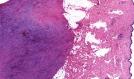
lesions. Microscopic examination: Subtypes: - Fascial type- Poorly circumscribed lesion , extends along the superficial fascia and interlobular septa of subcutaneous fat. - Subcutaneous type - Well circumscribed lesion, extends into the subcutis. - Intramuscular type - Well circumscribed lesion , grows into the skeletal muscle. - Intradermal type - Lesion present in the dermis (intradermal fasciitis). Early cases of Nodular fasciitis display zonation effect with maturation from the centre (hypocellular or hyalinized) to the periphery (hypercellular with inflammatory cell , blood vessels). In between, the loose myxoid area is populated by non- pleomorphic myofibroblasts loosely arranged with a tissue culture look . The backround stroma shows variable myxoid change. Extravasated red blood
cells and lymphocytes (not plasma cells) are also present. Hyalinization and keloidal change may be noted in longstanding cases. There are 1-2 normal mitotic figures per 5 / HPF (Note: More than 1 mitosis / HPF and atypical forms raises the possibility of a malignant tumour). Immunohistochemistry : NF demonstrates focal smooth muscle and muscle specific actin and calponin, but not usually desmin, h-caldesmon or CD34. CD68 may be positive in some cases. Similar microscopic features are present in reactive fasciitis-like lesions occuring. - In deep soft tissue location,
Example- Nerve, parotid sheath . Example: (i) Postoperative spindle cell nodule - bladder, prostate, vagina. (ii) Inflammatory fibromyxoid tumour - bladder (iii) Proliferative funiculitis - spermatic cord Differential diagnosis: A) Benign tumours: 1.Benign fibrous histiocytoma- Classical - Epidermal hyperplasia, peripheral collagen bundles, foamy macrophages and Touton giant cells. Cellular variant- Fascicular spindle cell architecture. 2. Neurofibroma- Architecture is different, S100 protein is positive. 3. Spindle cell lipoma - Fat, ropy collagen, absence of markers
4. Fibromatosis- More infiltrative growth pattern, slender spindle shaped
fibroblasts arranged in sweeping fascicles and separated by abundant
intercellular collagen. 1. Leiomyosarcoma- The cells in fasciitis are tapered and the nuclei are tapered rather than blunt ended. Atypical mitotic figures are prominent. Immunohistochemistry reveals h-caldesmon and desmin positivity. 2. Low grade myofibrosarcoma (myofibroblastic sarcoma) shows focal nuclear atypia,less inflammation, more uniformly cellular, reaches a larger size and infiltrates muscle. 3. Inflammatory myofibroblastic tumour has fasciitis-like,fascicular and fibrous areas and a marked plasma cell infiltrate. Immunohistochemistry reveals that some cases are cytokeratin and ALK-1 positive. 4. Myxofibrosarcoma is multinodular, has vacuolated fibroblasts and shows nuclear pleomorphism, abnormal mitosis, distinct vascular pattern and is usually actin negative (some are CD34 positive). 5. Malignant peripheral nerve sheath tumour has alternating cellular and myxoid fascicles, is more uniform and has wavy buckled and bullet shaped nuclei. Better differentiated case are at least focally
S100 protein positive and myoid markers are negative. 1) Absence of atypia 2) Absence of atypical mitotic figures 3) Small size 4) Short history 5) Superficial location in young adults. 1) Ossifying fasciitis: Nodular fasciitis like fibroblastic proliferations with metaplastic bone formation. 2) Intravascular fasciitis: Involve small or medium-sized veins or arteries. Histologically the features are similar to nodular fasciitis, however there are greater number of multinucleate giant cells and less prominent mucoid matrix. 3) Cranial fasciitis: The lesion involves the soft tissue of the scalp land is usually present in infants. Histologically this is well circumscribed lesion showing NF like fibroblastic proliferation in a prominent myxoid stroma.
|
|
|
|
|
