Custom Search

Dermpath-India

Pathology of Trichoepithelioma

Dr Sampurna Roy MD 


Dermatopathology Quiz Case: 163

Answer -  Trichoepithelioma


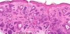
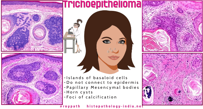
Trichoepithelioma is regarded as a poorly differentiated hamartoma of the hair germ. 

The tumour has three clinical forms: solitary, multiple, or desmoplastic.

Related post: Desmoplastic trichoepithelioma

Although most classifications include this as a separate entity Ackerman and his colleagues have suggested that such tumours should be grouped together with trichoblastomas and use the term "trichoblastoma" for all tumours showing predominant hair germ differentiation.

Site:  Commonly located on the head and neck region.

Solitary- Nose, upper lip and cheeks.

Multiple- Central part of the face, trunk, neck and scalp.

Clinical presentation: Skin coloured papules. May be solitary or multiple.

Papules may coalesce to form plaques. Rare presentation is a linear form.

Multiple tumours (epithelioma adenoides cysticum) inherited as autosomal dominant.

Brooke-Spiegler syndrome: Multiple trichoepitheliomas and Cylindromas .

Visit:Brooke-Spiegler Syndrome-[Pathology Infographic]


Microscopic features :

Well circumscribed dermal tumour ;

Islands,nests and cords of uniform basaloid cells ;

Cells are set in a variably cellular fibrous stroma ;

Epithelial structures resemble hair papillae or abortive hair follicle ;

Small keratocysts (infundibular differentiation) lined by stratified squamous epithelium ;

Retraction of stroma from adjacent dermis ;

Foci of calcification are often present ;

Few mitotic figures and apoptotic bodies.


Immature trichoepithelioma: 

Typically exhibits no horn cysts, displays fewer primitive hair structures, and lacks the adenoidal growth patterns of the tumour lobules which are usually present in the classical trichoepitheliomas.

Differential diagnosis :

1. Basal Cell Carcinoma:  

Distinction from some types of basal cell carcinoma has been based on the paucity of mitoses and apoptotic bodies, lack of retraction between stroma and epithelium and presence of primitive follicles in trichoepithelioma and differences in CD34 and bcl2 expression.

However, there are some tumours in which this distinction cannot be reliably made.   

2. Microcystic Adnexal Carcinoma ; 

3. Trichoadenoma

4. Trichoblastoma ;


Dermatopathology Quiz Case 12


Further reading:

Familial Facial disfigurement in Multiple Familial Trichoepithelioma.

Multiple trichoepitheliomas: A rare occurrence.

A clinicopathologic and molecular biologic study of patients presenting with few adnexal tumors (two to four) from the morphological spectrum of Brooke-Spiegler syndrome.

Value of CD10 Expression in Differentiating Cutaneous Basal from Squamous Cell Carcinomas and Basal Cell Carcinoma from Trichoepithelioma.

CD10 expression helps to differentiate basal cell carcinoma from trichoepithelioma.

Differentiation between basal cell carcinoma and trichoepithelioma by immunohistochemical staining of the androgen receptor: an overview.

The cytology of giant solitary trichoepithelioma.

Multiple familial trichoepithelioma: a case report and review of literature.

p75 Neurotrophin receptor differentiates between morphoeic basal cell carcinoma and desmoplastic trichoepithelioma: insights into the histogenesis of adnexal tumours based on embryology and hair follicle biology.

Multiple trichoepitheliomas associated with a novel heterozygous mutation in the CYLD gene as an adjunct to the histopathological diagnosis.

Diagnostic utility of immunohistochemistry in distinguishing trichoepithelioma and basal cell carcinoma: evaluation using tissue microarray samples.

A novel missense mutation of CYLD gene in a Chinese family with multiple familial trichoepithelioma.


Visit:- Infectious Disease Online

Dr Sampurna Roy  MD

Consultant  Histopathologist (Kolkata - India)


Copyright © 2022  histopathology-india.net