Custom Search
|
|
Custom Search
|
|
Dermpath-India Pathology of Pleomorphic Fibroma Dr Sampurna Roy MD 2022
|
|
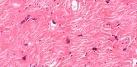
Pleomorphic Fibroma of the skin is a rare cutaneous fibrous tumor first described by Kamino et al in 1989. Clinically presents as a slow-growing polypoid skin tag-like or dome shaped lesion in middle aged and elderly patients. Site: On the trunk or extremities and rarely in head, neck, and subungual locations. Microscopic features: Well-circumscribed tumour composed of pleomorphic mononucleated and multinucleated cells with atypical nuclei with rare mitotic figures. The cytologic pleomorphism is considered as a degenerative process as seen in a number of other mesenchymal tumours. Immunohistochemistry: CD34 positive. Desmin, Ki-M1p antibodies (pan-monocytic/macrophage marker) or S100 protein-Negative. Staining for factor XIIIa has been patchy. Differential diagnosis: Benign tumours: Dermatofibroma with atypical cells ; Giant cell fibroblastoma, pleomorphic lipoma ; sclerotic fibroma. Malignant tumours: Undifferentiated pleomorphic sarcoma ; Atypical fibroxanthoma; Limited local recurrence may occur but there have been no reports of regional extension or metastatic spread.
|
|
|
 |
Visit:- Infectious Disease Online
![]()
Copyright © 2002-2022 histopathology-india.net