Custom Search
|


|
Dermpath-India Pathology of Angioleiomyoma Dr Sampurna Roy MD 2023
|
 |
|
Angioleiomyoma are
common typically painful,benign
neoplasm that originates from smooth muscle cells of arterial or venous
walls and contains thick-walled vessels. Site: These are usually located in the subcutis and deep dermis of the extremities, particularly the lower leg. Cases have been reported on the the head and upper trunk and these are often painless. Cases have been reported at unusual sites: i) Oral angioleiomyomas are usually located on the lip, palate, buccal mucosa and tongue, mandible, floor of mouth, and gingiva. ii) Tonsil iii) Auricle iv) Within the scrotum, structures such as the epididymis, spermatic cord, dartos fascia, and blood vessels could be sites of origin of angioleiomyoma. Macroscopic features: Presents as a slow growing , firm, gray white, round to oval nodules (usually less than 2cm in diameter). Age: Usually occur in the fourth and sixth decade of life.
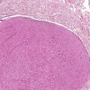
Microscopic features: Image1 ; Image2 ; Image3 ; Image4 ; Image5 Well circumscribed and encapsulated tumour is usually noted in the lower dermis and subcutis. There are interlacing bundles of smooth muscle fibres between vascular channels. The vessel walls (veins) display layers of smooth muscle fibres. These muscle fibres appear to merge peripherally into the intervascular muscle fibres. These vessels may have slit-like or dilated lumen. Degenerative changes may be present including vascular thrombosis, stromal hyalinization, myxoid changes, dystrophic calcification and nuclear atypia. Adipocytes may be
present in a few cases (also known as
angiomyolipoma, these are HMB45
negative unlike renal angiomyolipomas). Variants of Angioleiomyomas: 1. Solid; 2. Cavernous; 3. Venous ; 4. Epithelioid ; 5.Pleomorphic Note: Angioleiomyoma is often associated with pain. Pain is probably mediated by nerves present in the tumour and in the capsule due to either mechanical stretching or through mast cell mediation. Other painful tumours of soft tissue and skin are glomus tumour, spiradenoma, angiolipoma and traumatic neuroma.
|
|
|
|
|