

Custom Search
|


|
Dermpath-India Pathology of Calcifying Aponeurotic Fibroma Dr Sampurna Roy MD 2022
|
 |
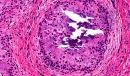
Calcifying aponeurotic fibroma was described as 'Juvenile aponeurotic fibroma' by Keasby in 1953. It is a rare, locally aggressive fibroblastic lesion located in the palms of the hands and soles of the feet in young children. These lesions have a recurrence rate of greater than 50% following surgical resection. Age and sex: Male patients are twice as commonly affected as female patients, and most of the lesions occur within the first two decades of life. Clinical presentation: The lesion presents as a slow-growing painless soft tissue mass on the hands or feet of several months or even years duration. Site: The lesions are mainly located in the hands or feet. In the hand most of the lesions occurred in the palm or the fingers. In the foot most of the lesions are located in the plantar or ankle region. Cases have also been reported in the toes. Some of the lesions are located at other sites including the forearm, elbow, popliteal fossa and supraclavicular region. Macroscopic features: Poorly defined, firm or rubbery, grayish white mass. The lesions are often adherent to dense fibrous connective tissue (Example: tendon, fascia, or periosteum) and ranged from 1.0 to 5.0 cm in maximum dimension. Focal punctate calcific areas may be present. This may give a gritty sensation during cutting of the specimen. Microscopic features: Biopsy is important in evaluation of these lesions. Microscopic examination showed spindled fibroblasts with a fascicular growth pattern and scattered epithelioid cells bordering chondroid foci with or without mineralization. The plump spindle cells contain round or ovoid nuclei and indistinctly outlined cytoplasm. There is a linear or palisading arrangement specially around calcified material. Some of these cells resemble chondrocytes. Occasionally osteoclast-like giant cells are present. There are no atypical features and mitoses are scarce. Backround stroma is densely collagenous. Calcifying aponeurotic fibroma may not exhibit foci of calcification in its earliest phase and often infiltrates fat and striated muscle at the periphery in infants and young children. Calcification and cartilage formation are more prominent in older children and young adults. Immunohistochemistry: Muscle-specific actin (+/-), smooth muscle actin (+/-), CD99 (+), S100 protein (+), CD68 (+). Differential diagnosis: Rheumatoid nodule ; Chondroma of soft parts; Fibrous hamartoma of infancy; Schwannoma; Fibromatosis. Calcifying Aponeurotic Fibroma – A rare, benign fibrous tumour [Pathology Infographic]
|
|
|
|
|
