

Custom Search
|

|
Infectious Disease Online Pathology of Cytomegalovirus Infection
|
|
Cytomegalovirus is a member of
the
Herpesviruses
and contain double-stranded
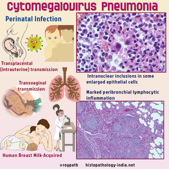
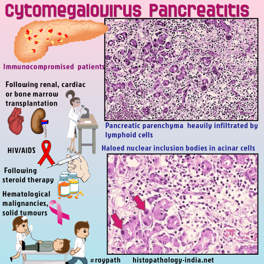
DNA. Description of the organism: Characterized by intra-nuclear inclusion with enlargement of both cell & nucleus. Cytoplasmic inclusions are also seen in the infected cells. Nuclear inclusion: Single, measuring up to 20 µm in diameter, while the cytoplasmic inclusions are much smaller, measuring 1 to 3 µm. It is usually basophilic but may be eosinophilic, round to oval, with a peripheral halo and accentuation of the nuclear membrane. At the edge of the viral inclusion, a rounded clump of peripheral chromatin may be seen extending into the clear region of the halo. Following Ganciclovir therapy, the intranuclear inclusion of Cytomegalovirus has been reported to have a globular and eosinophilic appearance due to loss of viral DNA. Cytoplasmic inclusions: Basophilic to amphophilic and indistinct. Special stain:Cytoplasmic inclusions stain with the Periodic Acid-Schiff (PAS) and Gomori-Methenamine Silver (GMS) stains. The intranuclear inclusions are Feulgen positive and contain viral nuclear, protein, and capsid material. Cases in which the inclusions are not easy to recognize or few in number and it is helpful to perform either immunohistochemical staining or in situ hybridization as a diagnostic aid. Epidemiology: It is common all over the world. 40-100% adult population is sero-positive. Transmission: 1. From mother: i) In-uterus (transplacental) ; ii) birth canal secretion-during birth. iii) breast milk 2. From Day-Care-Center through saliva. 3. Commonly from venereal contact, respiratory secretion & fecal-oral route. 4. Iatrogenic- (i) Blood transfusion ; (ii) Transplant from infected donor ; (iii) Congenital Cytomegalovirus infection. Mostly asymptomatic, Cytomegalovirus remains latent in leucocytes (major reservoir). Some produce fatal syndrome, known as Cytomegalic Inclusion Disease (CID). Clinical presentation: Jaundice, petechiae, microcephaly, thrombocytopenea, diarrhea and Central Nervous System disease. Pathology: Liver: Clinicopathological study of liver involvement in cytomegalovirus infection in infant autopsy cases. i) Persistence of hepatic hematopoesis. ; ii )Periportal hepatic necrosis iii)Dense mononuclear infiltrate. Adult Cytomegalovirus Infection: Mostly in immunosuppressed individuals. Example: i) Organ transplant from seropositive donor associated with immunosuppressive therapy. ii) Bone marrow transplant-due to immunosuppressive therapy & Graft vs Host reaction. iii) AIDS patients primarily affect Lungs & Gastrointestinal tract. Lung: Cytomegalovirus can infect a variety of cell types and in the lung - Epithelial cells of the airways and alveoli ; fibroblasts ; macrophages ; endothelial cells. The cytopathic effects of CMV include both nuclear and cytoplasmic inclusions. Infected cells show cytomegaly or marked enlargement. Four major patterns of Cytomegalovirus infection of the lung : i) Miliary pattern: Result from hematogenous spread ; shows multicentric lesions ; consist of alveolar exudation by fibrin, inflammatory cells, neutrophils, and chronic inflammatory cells. Centrally - Lesions may show necrosis, hemorrhage, alveolar fibrin, and inflammatory cells. Cytomegalovirus inclusions are present in the nodular lesions. ii) Diffuse interstitial pneumonitis: Features range from mild to a pattern of diffuse alveolar damage. There may be interstitial edema, alveolar fibrin, and hyaline membranes, or interstitial fibroblastic proliferation and type 2 pneumocyte hyperplasia. Number of Cytomegalovirus inclusions vary from a few to numerous. iii) Hemorrhagic pneumonia iv) Cytomegalovirus inclusions associated with minimal inflammation or lung injury: Cytomegalovirus pneumonia frequently occurs in immunocompromised patients. Combined infection of lung with Cytomegalovirus, is commonly seen with Pneumocystis carnii , Candida , Aspergillus , Nocardia and Mycobacteium Tuberculosis. Special techniques help to confirm the presence of multiple organisms, especially if more than one virus is present .
Gastrointestinal tract: - PubMed links to related abstracts In Cytomegalovirus infection of Gastrointestinal tract patient presents with severe pain, loss of weight, weakness, remitting fever. Gastrointestinal lesions are erosive-ulcerous or ulceronecrotic. Infection and viral replication, mostly occurs in ulcerative disease of intestine. Example: Crohns’ disease, Ulcerative colitis. Inclusions are seen in endothelial & mucosal cells. Pathogenesis of Cytomegalovirus infection in the gastrointestinal tract: The following pathogenetic chain of Cytomegalovirus infection course in the gastrointestinal tract was established: Vasculitis -> microcirculatory disorders -> segmental ischemia -> necrosis with inflammatory infiltration and CMV transformation of the cells-> fibrosing -> cicatricial transformation of the organ wall. Some authors have suggested that developing sclerosis due to cytomegalovirus involvement of the intestine may promote cancer. Skin: Few cases of cutaneous involvement have been reported: Microscopic features: i) Non-specific dermal infiltrate ; ii) Cells involved include - Endothelial cells in small dermal vessels ; fibrocytes ; macrophages and rarely ductal epithelial cells. 'Blueberry muffin lesions' seen in congenital infections are due to dermal erythropoiesis. Diagnosis: 1. Demonstration of CMV ; 2. Antiviral antibody ; 3. PCR- detection of viral genome.
|
|
PUBMED: Articles related to Cytomegalovirus Infection
|
|
|
