

Custom Search
|

|
Infectious Disease Online Pathology of Cat Scratch Disease
|
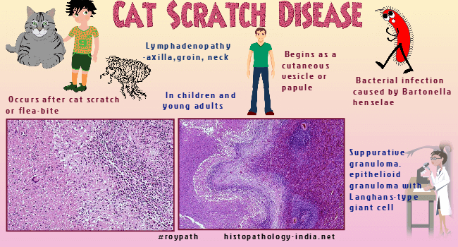
Cat scratch disease (CSD) is usually a
self-limited infection caused by a curved gram-negative, bacteria
Bartonella
henselae. The bacteria form filaments up to 10 micrometer or longer.It is easily seen in tissue sections of the skin, lymph nodes, and conjunctiva, when stained by a silver impregnation technique.Cats are the principal reservoir of Bartonella henselae, the etiologic agent in most cases of Cat scratch disease.Infection begins when the organism is inoculated into the skin by the claws of cats and rarely by other animals, or by thorns or splinters.Sometimes the conjunctiva is contaminated by close contact with a cat, possibly by licking around the eye.Infections are more common in children (80%) than in adults.Most patients have a papule at the site of inoculation. These may be small and can be easily overlooked. The papule, which begins 3 to 14 days after inoculation may persist for 8 weeks, is followed by tenderness and enlargement of the regional lymph nodes. The nodes remain enlarged for 3 to 4 months and may drain through the skin. About one-half of the patients have other symptoms, including fever and malaise and (rarely) splenomegaly, Parinaud’s oculoglandular syndrome, rash, encephalitis (which typically has a sudden onset and sudden resolution), and erythema nodosum. Rare complications of Bartonella henselae infection is bacillary angiomatosis. At the site of inoculation the bacteria multiply in the wall of the small vessels and about collagen fibers from which they move through draining lymphatics to regional lymphnodes, where they produce a pyogranulomatous lymphadenitis. In early lesions clusters of bacteria expand and obliterate the walls of small vessels. The lesions in the skin and lymphnodes progress from abscesses to suppurating granulomas and finally to necrosis. Bacteria are abundant in early lesions and rare in late ones. Without biopsy and the visualization of the characteristic bacteria, the diagnosis is supported when three criteria are met : (i) contact with a cat, a cat scratch, or a primary lesion of the skin or conjunctiva. (ii) A positive skin test for cat scratch antigen and (iii) Negative results from laboratory studies for other causes of lymphadenopathy. Although serologic testing is the reference method for diagnosis, successful use of immunohistochemical (IHC) stain of regional lymph nodes for the diagnosis of Cat scratch disease has been reported. The histologic differential diagnosis of CSD lymphadenitis includes Kikuchi necrotizing lymphadenitis , Kawasaki disease and other infectious processes, such as tularemia, mycobacterial infection, brucellosis, fungal infection, lymphogranuloma venereum and lymphadenitis associated with idiopathic granulomatous mastitis (especially when in a young female patient). Lymphogranuloma venereum mainly involves the inguinal lymph node. Serologic studies, special stains for Bartonella, and molecular studies might be required to distinguish between the two. Related posts: Bartonellosis ; Verruga peruana ; Bacillary angiomatosis. |
|
|
