

Custom Search
|

|
Infectious Disease Online Pathology of Verruga Peruana - Bartonellosis
|
|
Syn: Chronic Carrion's disease
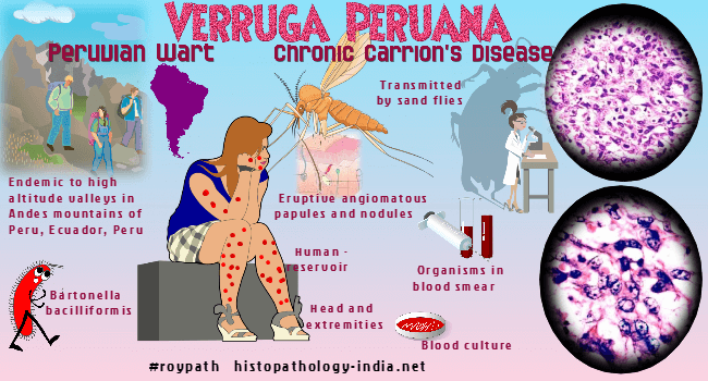
; Peruvian Wart Bartonella bacilliformis, a gram-negative, flagellated,motile bacterium, is the etiologic agent of Carrion's disease, the chronic cutaneous form of which (verruga peruana), presents as superficial and deep angiomatous cutaneous nodules. Related post: Bartonellosis ; cat-scratch disease ; bacillary angiomatosis. Carrion's disease, trench fever, cat-scratch disease, and bacillary angiomatosis are examples of Bartonella sp. infection. Bartonellosis or Carrion's disease is endemic in some regions of Peru, Ecuador and Columbia. The foci of endemic transmission are river valleys in the Andes between 500 and 3200 meters above sea level. The disease is transmitted by the sandflies Phlebotomus verrucarum and Phlebotomus noguchi. Humans provide the only reservoir and acquire bartonellosis at sunrise and sunset, when sandflies are most active. In endemic areas, 10% to 15% of the population have latent infection. Newcomers are susceptible whereas indigenous population is resistant due to subclinical infection and and immunity. Clinical presentation: There are two syndromes both caused by B. Bacilliformis - Oroya fever (acute anemic phase) and verruga peruana (the chronic dermal phase). These present as a biphasic pattern with the acute anemia first followed some months later by the chronic dermal phase. Occasionally, the dermal eruptive form develops independently without prior evidence of bartonellosis. Skin lesion: 1. May present as multiple, miliary, superficial, small hemangioma like-lesions of the dermis which balloon outward and cause a studded appearance. 2. Nodular lesions are larger but fewer and may be more prominent on the extensor surfaces of the arms and legs. Sometimes large deep seated lesions which tend to ulcerate, develop near joints and limit motion. Histopathology: Histologically, specimens of all the verruga nodules has features consistent with granulomatous lesions with extensive infiltration of various types of cells along with the proliferation of capillary-like vessels in the papillary dermis with the formation of a collarette. The sections are predominantly infiltrated with neutrophils and endothelial cells. Histiocytes, plasma cells, lymphocytes and mast cells are also visible. The blood vessels are dilated, and many rounded and swollen endothelial cells are located peripherally. A huge number of neutrophils invaded the vessels.
|
|
|

![]()
Copyright © 2022 histopathology-india.net