

Custom Search
|


|
Dermpath-India Pathology of Hibernoma Dr Sampurna Roy MD 2022
|
|
Hibernomas are rare, asymptomatic,
benign tumours that arise mostly in adults from the remnants of
fetal brown adipose tissue, and usually affect muscle and
subcutaneous tissue.
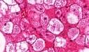
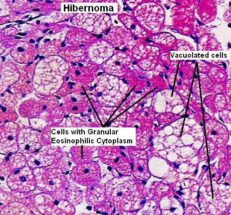
This tumor was first described by Merkl in 1906 [Uber ein Pseudolipoma der Mamma (elgenartiger Fettzellentumor). Beitr Pathol Anat Allg Pathol 1906;39:152–7]. The brown fat, described by Velch in 1670, is a specialized form of fat found in the hibernating and non hibernating animals such as rats, cats, monkeys, rabbits, and humans. Human newborns have high levels of brown fat. Age: Usually occurs between ages 20 and 50 years. Site: In the adult, brown fat is usually found in scattered foci as persisting vestigial remnants along the oesophagus, trachea, posterior neck, and interscapular area and around the great vessels of the mediastinum. Hibernomas are usually seen at one of these sites. The most common anatomic locations included the thigh, shoulder, back , neck, chest, arm and abdominal cavity/retroperitoneum. Gross: The tumour has been described as well encapsulated, tan-brown lobulated tumour. However, infiltration of adjacent structures, particularly striated muscle may be present. Microscopic features: The Hibernoma is composed of so-called mulberry cells with scanty stroma. The characteristic mulberry cell has a centrally located nucleus and multivaculoated foamy cytoplasm in which eosinophilic fat droplet granules are present. Four morphologic variants of hibernoma were identified: typical ; myxoid ; spindle cell, and lipoma-like. 1. Typical hibernoma included eosinophilic cell, pale cell, and mixed cell types based on the tinctorial quality of the hibernoma cells. 2. The myxoid variant contained a loose basophilic matrix. 3. Spindle cell hibernoma had features of spindle cell lipoma and hibernoma. All occurred in the neck or scalp. 4. The lipoma-like variant contained only scattered hibernoma cells. General features: The tumour is divided into lobules by thin septa: There are three cell types: i) Large coarsely vacuolated cells ii) Large finely vacuolated cells with eosinophilic granular cytoplasm. iii) Mature univacuolated adipocytes. Vacuoles stain positively with oil red O in frozen section. Immunohistochemistry: S100 protein- Positive. Positivity may be focal to diffuse. In some cases (spindle cell variant) CD34 may be positive. Cytokeratin, EMA and CEA are negative. Cytogenetic: Rearrangements of 11q13 (may result in deletion of MEN-1 gene locus). Abstract Differential diagnosis: Lipoma: In cases with numerous univacuolar cells, the differential diagnosis from lipoma may be difficult but the ultrastructural features are different for both types of tumors. Adult rhabdomyoma : Made up of similar eosinophilic cells but its cells are larger and contain glycogen. Cross-striations are seen . Chondroid lipoma : Contains round or polygonal eosinophilic cells, many with lipid vacuoles closely simulating lipoblast and it shows features of both lipoma and hibernoma, but the stroma contains chondroid material which is not present in the hibernoma. Liposarcoma : Myxoid and round cell liposarcomas may contain hibernoma-like cells. However, large numbers of hibernoma cells are absent and mitotic activity is prominent. Granular cell tumour : Distinguished by complete absence of lipid cytoplasmic vacuoles. The granular cells are diffusely positive with S-100 protein. Metastatic renal cell carcinoma: Immunohistochemistry helps in distinction (Cytokeratin, EMA and vimentin positive). Treatment : Although hibernomas are considered benign, they tend to enlarge in size sometimes causing compression of the neighbouring structures. Complete excision is the treatment of choice.
|
|
|
 |
