

Custom Search
|


|
Dermpath-India Lichen Planus-like Cutaneous Lesions Dr Sampurna Roy MD 2022
|
| Pathology of Lichen
Planus
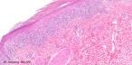
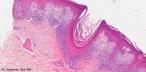
Lichen planus is an inflammatory disorder of the skin and mucous membranes with no known cause. It appears as pruritic, violaceous papules and plaques most commonly found on the wrists, lower back, and ankles.Lichen planus was first described by Erasmus Wilson in 1869. Microscopic features: Orthokeratoic hyperkeratosis, wedge shaped hypergranulosis and saw-tooth appearance of the epidermis; Effacement of the dermoepidermal junction by band-like mononuclear inflammatory cell infiltrate (interface dermatitis) ; Hydropic degeneration of basal cells ; Colloid body formation ; Pigment incontinence; Variants of Lichen Planus: Atrophic Lichen Planus: Loss of normal rete ridges. Inflammatory infiltrate less dense. Hypertrophic Lichen Planus: Epidermal hyperplasia with changes limited to the tips of the rete-ridges. Often superadded lichen simplex chronicus. Ulcerative Lichen planus: Common sites include feet, perineum, vulva, vagina and mouth. Changes present at the edge of the ulcer. Plasma cells are present in the mucosal lesions. Erythema Dyschromicum Perstans: Macular variant of lichen planus. Prominent melanin incontinence. Lichen Planus Actinicus: Usually in young individuals of Oriental origin. Prominent melanin incontinence. Lichen Planus Pemphigoides: Cell poor subepidermal bulla. Lichen Planopilaris: Infiltrate extends around hair follicle. Lichenoid keratosis (lichen planus-like keratosis): Presents as solitary lesion on sun-exposed skin. Site: Usually located on the upper limbs. Microscopic features: Histopathological features mimic lichen planus. Prominent Civatte body formation. Serial sectioning reveals solar lentigo at the margins. Focal parakeratosis (+). Hypergranulosis not as prominent as in lichen planus. Lichenoid Drug Eruption: Microscopic features: Focal parakeratosis, spongiosis and mild basal cell degeneration. Some eosinophils and plasma cells in the dermal infiltrate. Inflammatory infiltrate extends around blood vessels in the mid and deep dermis. Sometimes drug eruptions may present with a hypertrophic lichen planus- like picture. Rarely multinucleated giant cells are present known as 'giant cell lichenoid dermatitis'. Fixed Drug Eruption: Microscopic features: Lichenoid reaction pattern, hydropic degeneration and presence of necrotic keratinocytes in the basal layer and higher up in the epidermis. Prominent melanin incontinence is present. Inflammatory infiltrate obscure the dermoepidermal junction. Extends upto mid or upper epidermis. Lichenoid Graft Versus Host Disease: Cutaneous lesion in graft-versus host disease Clinical history is important. Microscopic features: Inflammatory cellular infiltrate is not band-like and is less prominent than in lichen planus. Necrotic keratinocytes present at all levels of epidermis , accompanied by lymphocytes (satellite cell necrosis). Lichen Striatus: Microscopic features: Features may mimic lichen planus. However, interphase changes are focal (occupy three or four adjacent dermal papillae). There is focal spongiosis. Deep perieccrine inflammatory infiltrate is present. Other lesions showing lichen planus-like (lichenoid) reaction: Lichenoid reaction to tumour following regression of melanocytic or epithelial tumours. In a tattoo there are macrophages containing tattoo pigments.
Discoid Lupus Erythematosus: Pathology of Discoid Lupus Erythematosus Well demarcated erythematous scaly patches. Site: Face, cheek, bridge of nose, sometimes neck, scalp, lips, oral mucosa. Microscopic features: Hyperkeratosis and follicular plugging. Atrophy of epidermis. Lichenoid reaction pattern characterized by vacuolar degeneration and presence of Civatte bodies. Thickening of the basal membrane. Superficial and deep perivascular and periadnexal inflammatory infiltrate.
|
|
|

![]()
Copyright © 2022 histopathology-india.net