

Custom Search
|

|
Infectious Disease Online Pathology of Orf (Ecthyma Contagiosum) Dr Sampurna Roy MD 2022
|
|
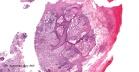
Orf virus is a DNA parapoxvirus that causes orf (ecthyma contagiosum) , an acute debilitating skin disease of sheep, goats and humans. This double-stranded DNA poxvirus is a member of the Paravaccinia subgroup. Orf (ecthyma contagiosum) usually causes crusted lesions on the lips and perioral area. The first case of human orf was documented in 1934 by Newson and colleagues. Visit: Variola (smallpox) ; Molluscum Contagiosum Mode of infection: Orf can be transmitted to humans by contact with infected animals. In some cases the lesion developed at sites of trauma produced by an inanimate object. In most instances, the human disease occurs among sheep handlers and is transmitted by direct contact with the papulovesicular lesions or the crusts from an infected animal Site: It occurs most commonly on the index finger. The lesions (about 1-3 cm or more in diameter) are usually located on the hands and forearms. Other sites include the face, scalp, temple, and perianal region. Clinical presentation: Several lesions may be present in the one area. A mature lesion is nodular with central umbilication and an erythematous halo. Complications: The characteristic lesion resembles a tumour and resolves spontaneously (after about 7 weeks) , usually without any complications. However, rare complications such as lymphangitis, adenitis, erythema multiforme, toxic erythema , erysipelas, papulovesicular eruption . Pseudomonas aeruginosa infection, and bullous pemphigoid have been reported. In one case giant orf caused swan-neck deformity and paresthesia. Recurrent lesions have been reported in immunocompromised persons. The features depend on the stage of the disease. Early lesions : - Moderate epidermal acanthosis ; - Pale vacuolated cytoplasm, mainly in the upper epidermis ; - Cytoplasmic inclusion bodies are usually present; - "Spongiform degeneration" - vacuolated cells having wispy strands of eosinophilic cytoplasm. Usually seen in follicular structures. - Intraepidermal vesicles or bullae may form. - Dilated thin-walled vessels and an infiltrate of lymphocytes, macrophages and occasional eosinophils and plasma cells in the dermis Later lesions: - Epidermal necrosis, mainly in the center. - Neutrophils are often present within and close to the necrotic epidermis. - Other biopsies may show elongated rete pegs with dilated vessels in the intervening dermal papillae. - Sometimes there is an unusual proliferation of endothelial cells in the dermal papillae. This feature may resemble a vascular tumour. - The inflammatory infiltrate and epithelial hyperplasia finally resolve. Differential diagnosis: Cowpox and smallpox virus infection : These lesions like orf may show multilocular vesicles and prominent ballooning of keratinocytes with cytoplasmic eosinophilic inclusion bodies. Should be differentiated from parapoxvirus infection by electron microscopy, virology, or PCR. Herpes simplex, varicella, and zoster - Multinucleated epithelial giant cells and keratinocytes with distinct chromatin margination and intranuclear eosinophilic inclusions are commonly present in these lesions. Molluscum contagiosum can easily be differentiated histologically from orf infection by the presence of cytoplasmic amphophilic bodies in the affected keratinocytes. Milker’s nodules : It is difficult to distinguish between the lesions of orf from milker’s nodules. Full-thickness epidermal necrosis seems to be more common in orf. Pyogenic granuloma diagnosis can be differentiated by the presence of a collarette of epidermis with pale edematous stroma. Pyoderma gangrenosum shows a mixed cellular infiltrate mainly composed of neutrophils and there is erythrocyte extravasation without mural necrosis or luminal fibrin deposition. Mycobacterium marinum of the skin can be differentiated by the presence of acid-fast bacilli and hyperkeratosis of epidermis, which are characteristic. Cutaneous anthrax should also be included in the histopathological differential diagnosis - gram stain reveals gram-positive rod-shaped bacilli. Diagnosis: Immunoperoxidase techniques using orf-specific monoclonal antibodies can be used to confirm the diagnosis. This zoonotic infection shares clinical manifestations and exposure risks with other, potentially life-threatening zoonoses (Example. cutaneous anthrax) and is likely under-recognized because of a lack of clinical suspicion and widely available diagnostics. Barrier precautions and proper hand hygiene are recommended for the prevention of orf virus infection in humans.
Secondary bacterial infection in patients infected with the orf virus should be aggressively treated.
|
|
|
