Custom Search
|


|
Custom Search
|
|
Dermpath-India Pathology of Acanthomas
|
|
Acanthomas are benign tumours of the epidermal keratinocytes.
|
| 1) Epidermolytic
Acanthoma 2) Acantholytic acanthoma 3) Melanoacanthoma The benign acanthomas are the benign tumours of epidermal keratinocytes. [ The benign acanthomas. Brownstein MH. J Cutan Pathol. 1985 Jun-Aug;12 (3-4):172-88. An important, yet neglected, problem in dermatopathology, is the evaluation of the benign acanthomas, the benign tumors of epidermal keratinocytes. The benign acanthomas may be simulated by lesions which are not benign (e.g. actinic keratosis), not tumors (e.g. normal plantar skin), or are not epidermal (e.g. dermatofibroma). In addition to normal (epidermoid) keratinization (e.g., seborrheic keratosis and related conditions), the variants of the benign acanthomas show a wide range of aberrant keratinization, including epidermolytic hyperkeratosis (epidermolytic acanthoma), dyskeratosis (warty dyskeratoma), acantholysis (acantholytic acanthoma), cornoid lamellation (porokeratosis), lichenoid hyperplasia (lichen planus-like keratosis), and absence of keratinization (clear cell acanthoma). ]
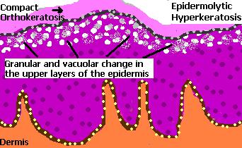
Epidermolytic
Acanthoma: A disseminated form has been described. Histology shows epidermolytic hyperkeratosis.
[ Epidermolytic hyperkeratosis is an abnormality of epidermal maturation. Features include compact hyperkeratosis, granular and vacuolar degeneration of the cells of the spinous and granular layers. ] Further reading: Solitary epidermolytic acanthoma. Epidermolytic acanthoma of the scrotum: A rare mimicker of condyloma acuminatum. Linear epidermolytic acanthoma or adult-onset verrucous epidermal nevus? Solitary epidermolytic acanthoma: a clinical and histopathological study.
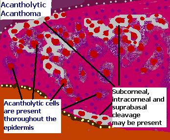
Acantholytic Acanthoma: This is a solitary tumour arising on the trunk of older patients . Multiple lesions have been reported in a renal transplant recipient.
Histology reveals hyperkeratosis, papillomatosis, acanthosis and variable acantholysis together with cleft formation. Further reading: Acantholytic tumor of the nail: acantholytic dyskeratotic acanthoma. Acantholytic dyskeratotic acanthoma: a variant of a benign keratosis.
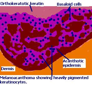
Melanoacanthoma:
Histologically, this lesion resembles a pigmented seborrheic keratosis. The features include acanthotic epidermis, composed of basaloid and spinous cells together with scattered dendritic melanocytes. Further reading: An unusual clinical presentation of gingival melanoacanthoma. Melanoacanthoma of external ear: report of two cases. Melanoacanthoma: Uncommon presentation of an uncommon condition. Oral melanoacanthoma: A rare case of diffuse oral pigmentation. Immunohistochemical features of multifocal melanoacanthoma in the hard palate: a case report. Oral melanoacanthoma: A rare case of diffuse oral pigmentation. Multifocal cutaneous melanoacanthoma with ulceration: a case report with review of literature. Multifocal oral melanoacanthoma and melanotic macula in a patient after dental implant surgery. Melanoacanthoma simulating pigmented spitz nevus: an unusual dermoscopy pitfall. |
|
|
 |
![]()
Copyright © 2002-2022 histopathology-india.net