Gastrointestinal Stromal Tumour


http://www.histopathology-india.net/SoftTissuePath.htm

                        HISTOPATHOLOGY INDIA.COM

         Atypical Fibroxanthoma 

            Dr Sampurna Roy  MD

 
Web www.histopathology-india.net
 May 2007

Surgical-Pathology.com

Histopathology-India.net

Eye Pathology Online

Cardiac Path Online;

Pulmonary Pathology Online

Pathology Quiz Online;

Dermpath-India;

GI Path Online

Mesothelioma-Online;

Soft Tissue Pathology;

Infectious Disease Online; INDEX: A-D ; INDEX: E-L ; INDEX: M-P INDEX: Q-Z ; FUNGAL DISEASE ; VIRAL DISEASE.

Nodular fasciitis

Proliferative fasciitis

Proliferative myositis

Ischaemic fasciitis

Elastofibroma

Fibrous Hamartoma of Infancy

Infantile Myofibromatosis/ Myofibroma

Juvenile hyaline fibromatosis

Inclusion  Body Fibromatosis

Calcifying aponeurotic fibroma

Fibromatosis colli

Fibroma of tendon sheath

Desmoplastic fibroblastoma

Storiform Collagenoma (sclerotic fibroma)

Giant Cell Collagenoma

Pleomorphic Fibroma

Angiomyofibroblastoma

Dermatomyofibroma

Cellular Angiofibroma

Fibromatosis

Lipofibromatosis

Solitary fibrous tumour

[Hemangiopericytoma  including Lipomatous Hemangiopericytoma]

Inflammatory myofibroblastic tumour

Low grade myofibroblastic sarcoma

Myxoinflammatory fibroblastic sarcoma

Infantile fibrosarcoma

Adult fibrosarcoma

Myxofibrosarcoma

Low grade fibromyxoid sarcoma

Hyalinizing Spindle Cell Tumour with Giant Rosettes

Sclerosing epithelioid fibrosarcoma

Fibrous Histiocytoma (Dermatofibroma)

Epithelioid Cell Histiocytoma 

Plexiform Fibrohistiocytic Tumour

Giant Cell Fibroblastoma

Dermatofibrosarcoma Protuberans  

Atypical Fibroxanthoma 

Malignant Fibrous Histiocytoma 

Adnexal Tumours of the eyelid

Mesenchymal Tumour of the Eyelid

Pulmonary Mesenchymal Tumours

Cardiac fibroma


Plexiform fibrohistiocytic tumour was first described by Enzinger and Zhang in 1988 and is characterized by fibrohistiocytic cytomorphology and a multinodular growth pattern.

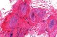
It is a rare mesenchymal neoplasm of children, adolescents, and young adults characterized by low-grade malignant behaviour. The tumour is prone to recur locally and occasionally to metastasize regionally and systemically.

The most common location is the upper extremities, followed by the lower extremities, trunk, head, and neck.

The tumour typically presents as a painless dermal or subcutaneous mass, which slowly grows for a period of months or years.

Gross features: Grossly, the tumour is usually described as being gray-white and firm and ranging in size from 0.5 to 8.0 cm.

Microscopic features: Microscopically, extensions of fibrous tissue into the surrounding fat identified on low-power is quite characteristic of Plexiform fibrohistiocytic tumour.

Most cases show a rim of normal dermis above the lesions and the overlying epidermis is unremarkable.

The nodular component consist of nests of mononuclear histiocytic cells that are often admixed with osteoclast-like multinucleated giant cells,  showing no nuclear atypia or necrosis . The clusters of mononuclear histiocytic cells are circumscribed by short fascicles of fibroblast-like cells that intersperse between collagen bundles.

Most tumours display mitotic activity, frequently <3 mitoses/10 high-power fields, and rarely the tumour may display atypical mitoses.

Based on proportion of fibroblast-like spindle cells and mononuclear histiocytic cells there are 3 histologic subtypes: i) fibrohistiocytic, ii) fibroblastic, iii) mixed.

The fibrohistiocytic type consists of nests of histiocytic cells interspersed with multinucleated giant cells.

The fibroblastic type consists of spindle fibroblast-like cells arranged in a plexiform pattern resembling fibromatosis.

There may be areas of haemorrhage which is a useful diagnostic feature.

Other rare features include granular cell change, hyalinization, osseous metaplasia and myxoid changes.

 Vascular invasion / intravascular growth has been observed in some cases.


Immunohistochemistry:

Vimentin-Positive ;  Alpha-1-antitrypsin & Alpha-1-antichymotrpsin- Positive

Smooth Muscle Actin - Positive (spindle fibroblast-like cells are commonly positive for smooth muscle actin and are occasionally positive for CD34).

CD68 -  stained mainly the multinucleated giant cells and, to a lesser extent, mononuclear histiocyte-like cells and, occasionally, fibroblast-like cells.

Desmin & S100 protein- Negative.

Electron microscopy:Show features of undifferentiated cells, myofibroblasts, and fibroblasts.

Differential Diagnosis: 

Dermatofibroma:   Older patients. Foam cells are present. No evidence of plexiform pattern and characteristic nodules present in Plexiform fibrohistiocytic tumour.

Tumours with a prominent plexiform pattern such as plexiform neurofibroma(S-100 positive, and usually associated with neurofibromatosis), plexiform schwannoma, fibrous hamartoma of infancy, cellular neurothekeoma, desmoid fibromatosis, and nodular fasciitis. 

Other tumours included in the differential diagnosis are atypical fibroxanthoma, pleomorphic MFH and giant cell sarcoma of soft part.

  Microscopic images of Plexiform fibrohistiocytic tumour:(ESCOP)                        Image Link1  2  3  4  5


Web www.histopathology-india.net
Abstracts:

Plexiform fibrohistiocytic tumor presenting in children and young adults. An analysis of 65 cases. Am J Surg Pathol. 1988 Nov;12(11):818-26.

Plexiform fibrohistiocytic tumor: clinicopathologic analysis of 22 cases. Am J Surg Pathol.1999;23(6):662-70

Dermal and subcutaneous variants of plexiform fibrohistiocytic tumor. Am J Surg Pathol. 1997 Feb;21(2):235-41

Plexiform fibrohistiocytic tumor. Eur J Dermatol. 2004 ;14(2):118-20

Plexiform fibrohistiocytic tumor without multinucleated giant cells. Am J Dermatopathol 2002;24:399–401.

Plexiform fibrohistiocytic tumor: a rare low-grade malignancy of children and young adults. Arch Otolaryngol Head Neck Surg 2002;128:966–970.

Myxoid plexiform fibrohistiocytic tumor. J Eur Acad Dermatol Venereol 2002;16:519–521.

Plexiform fibrohistiocytic tumor without multinucleated giant cells: a case report.Am J Dermatopathol. 2002;24(5):399-401

Plexiform fibrohistiocytic tumour - case report of a rare low-malignant tumour with frequent localisation at the upper limb.Handchir Mikrochir Plast Chir. 2002;34(3):201-4.

Plexiform fibrohistiocytic tumor: a rare low-grade malignancy of children and young adults. Arch Otolaryngol Head Neck Surg. 2002;128(8):966-70

Plexiform fibrohistiocytic tumor of soft tissues and bone.Cesk Patol. 2000;36(3):106-10

Fine needle aspiration cytology of plexiform fibrohistiocytic tumor. A case report. Acta Cytol. 1999;43(5):867-72

Fibrohistiocytic skin lesions.Verh Dtsch Ges Pathol. 1998;82:290-300

Plexiform fibrohistiocytic tumour with novel phenotypic features. Histopathology.1994 ; 25(2):165-9

Plexiform fibrohistiocytic tumor. Report of a case involving preoperative aspiration cytology and immunohistochemical and ultrastructural analysis of surgical specimens. Pathol Res Pract. 1992 ;188(3):350-6; discussion 356-9

       Your Banner
Pulmonary Pathology Online

Examination of pulmonary and pleural biopsies ; Percutaneous Needle and Trucut Biopsy Specimen  ; Bronchial Biopsy Specimen  ;Transbronchial Biopsy Specimen  ; Transbronchial biopsy in lung transplant recipients  ; Open lung biopsy  ; Lobectomy and pneumectomy specimen ; Histopathological reporting of pulmonary parenchymal biopsies ;Useful chromatic and immuno-stains in pulmonary pathology ;Congenital Cystic Adenomatoid Malformation ; Chondroid Hamartoma ; Acute Respiratory Distress Syndrome ; Neonatal Respiratory Distress Syndrome ; Complications of Neonatal Respiratory Distress Syndrome Extrinsic Allergic Alveolitis (Hypersensitivity Pneumonitis) ; Chronic Obstructive Pulmonary Disease ;Bronchial Asthma ; Bronchiectasis ; Chronic Bronchitis  ; Emphysema ;Bronchiolitis ; Lipid Pneumonia (Paraffinoma) ; Pulmonary Alveolar Proteinosis ;Pulmonary Thromboembolism ; Other forms of  Pulmonary Embolism ; Pulmonary Infarction ; Pulmonary Hypertension ; Pulmonary Collapse (Atelectasis) and Pneumothorax ; Pulmonary Edema ; Pulmonary Hemorrhage (Eg. Goodpasture's Syndrome) ; Sarcoidosis ; Lymphangioleiomyomatosis ; Localized Fibrous Tumour of the Pleura ; Pulmonary Lymphoproliferative Disease ; Lymphomatoid Granulomatosis ;Post-Transplant Lymphoproliferative Disease ;Biphasic Epithelial/ Mesenchymal Lung Tumours ;Pulmonary Carcinosarcoma ; Pulmonary Blastoma ;Large Cell Neuroendocrine tumour ;

Soft Tissue Pathology;

Myxoid Tumours of Soft Tissue Classification of Soft Tissue Tumour;  Gross examination of soft tissue specimen ;  A practical approach to histopathological reporting of soft tissue tumours Grading of soft tissue tumours ; Lipomatous tumours ;Neural tumours ; Myogenic tumours ;Vascular tumours ;Fibroblastic/Myofibroblastic tumours ; Myofibroblastic tumours ;  Fibrohistiocytic tumours ; ChondroOsseous tumours ; Soft TissueTumours of Uncertain Differentiation ; Notochordal Tumour -Chordoma ;Extra-adrenal Paraganglioma ; Gastrointestinal Stromal Tumour ;