

Custom Search
|


|
Dermpath-India Pathology of Epithelioid Sarcoma Dr Sampurna Roy MD 2022
|
|
Epithelioid sarcoma (ES) was first described
by Enzinger (1970 Epithelioid sarcoma. A
sarcoma simulating a granuloma or a carcinoma.Cancer 26:1029-1041).
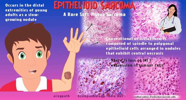
This rare malignant tumour occurs in adolescents and young adults. ES is usually located on the distal extremities (hands, fingers and forearm). This tumour rarely occurs on the pelvis, perineum and vulva. The tumour spreads along fascial and neurovascular structures with formation of satellite nodules. Multiple recurrences are known to occur in this tumour. Metastasis to the scalp, lymphnode and lungs occur frequently. Adequate treatment requires early radical excision or amputation if the tumour is located in the fingers or toes. Regional lymphnode dissection should be included as lymphnode metastasis is common in epithelioid sarcoma. Surgical excision is combined with radiotherapy. Loss of expression of INI1 is frequent in the conventional and large cell subtypes of ES and can be used as a diagnostic marker, but it has no prognostic impact.
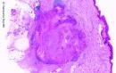
Gross features: The superficial lesion presents as a slow growing, firm tan white nodules or plaques, 0.5 - 5cm in diameter. These lesions are often ulcerated. The deeper lesions are attached to the tendon or fascia. These are larger in size and present as less well defined mutinodular masses or as an area of induration, usually 3-6 cm in diameter.
|
| Histological features: Usual Epithelioid Sarcoma - Distinct nodular appearance - Central necrosis of the tumour nodule. - Fusion of several necrotizing nodules with formation of 'geographic lesion'. - Tumour composed of oval or polygonal cells with eosinophilic cytoplasm. There is gradual transition into plump spindle cells. - Scattered mitotic figures. - Minimal nuclear pleomorphism. - Conspicuous inflammatory cell infiltrate composed of lymphocytes and histiocytes surrounding the tumour nodule. - Focal calcification and osseous metaplasia. - Intercellular dense hyalinized collagen. - In some cases cytoplasmic vacuolation may be present simulating lumen formation. These are intracellular lipid droplets. - The tumour may grow along neurovascular bundles and invest large vessels and nerves. Proximal -Type Epithelioid Sarcoma - Sheets of large epithelioid like cells with rhabdoid inclusions. - Central necrosis is absent - These are aggressive tumours and the differential diagnosis includes undifferentiated carcinoma and extrarenal rhabdoid tumour. Both classic and proximal-types are associated with the loss of SMARCB1/INI1 protein expression. Immunohistochemistry: The following markers are positive: Cytokeratin (K8 is positive in almost all cases, K19 is positive in some cases), EMA, CD34 (patchy membrane positivity), muscle specific actin (diffuse staining). Differential diagnosis:
(Tumour cells in ES are larger and more eosinophilic. Diagnosis is confirmed by immunohistochemistry) - Necrobiotic process - Granuloma annulare, Necrobiosis lipoidica, Rheumatic nodule
-
Dermatofibroma - Epithelioid vascular malignancy (Epithelioid angiosarcoma; Epithelioid hemangioendothelioma) - Melanoma - Squamous Cell Carcinoma (No dyskeratosis or any involvement of the epidermis in epithelioid sarcoma).
|
|
|

![]()
Copyright © 2022 histopathology-india.net